Light: Science & Applications ( IF 20.6 ) Pub Date : 2019-11-21 , DOI: 10.1038/s41377-019-0211-5 Biwei Yin , Zhonglie Piao , Kensuke Nishimiya , Chulho Hyun , Joseph A. Gardecki , Adam Mauskapf , Farouc A. Jaffer , Guillermo J. Tearney
|
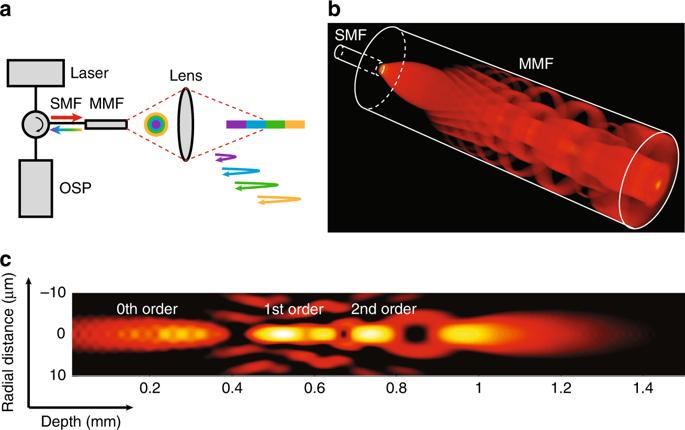
Cross-sectional visualisation of the cellular and subcellular structures of human atherosclerosis in vivo is significant, as this disease is fundamentally caused by abnormal processes that occur at this scale in a depth-dependent manner. However, due to the inherent resolution-depth of focus tradeoff of conventional focusing optics, today’s highest-resolution intravascular imaging technique, namely, optical coherence tomography (OCT), is unable to provide cross-sectional images at this resolution through a coronary catheter. Here, we introduce an intravascular imaging system and catheter based on few-mode interferometry, which overcomes the depth of focus limitation of conventional high-numerical-aperture objectives and enables three-dimensional cellular-resolution intravascular imaging in vivo by a submillimetre diameter, flexible catheter. Images of diseased cadaver human coronary arteries and living rabbit arteries were acquired with this device, showing clearly resolved cellular and subcellular structures within the artery wall, such as individual crystals, smooth muscle cells, and inflammatory cells. The capability of this technology to enable cellular-resolution, cross-sectional intravascular imaging will make it possible to study and diagnose human coronary disease with much greater precision in the future.
中文翻译:
使用少模干涉术的动脉3D细胞分辨率成像
体内人类动脉粥样硬化的细胞和亚细胞结构的横截面可视化非常重要,因为这种疾病从根本上说是由以深度依赖的方式在此规模下发生的异常过程引起的。然而,由于常规聚焦光学器件固有的分辨率深度深度折衷,因此当今最高分辨率的血管内成像技术,即光学相干断层扫描(OCT),无法通过冠状动脉导管以该分辨率提供横截面图像。在这里,我们介绍一种基于少模干涉法的血管内成像系统和导管,它克服了传统高数值孔径物镜的聚焦深度限制,并能够通过亚毫米直径,灵活的尺寸在体内进行三维细胞分辨率的血管内成像导管。用此设备采集了患病尸体的人冠状动脉和活体兔动脉的图像,显示出动脉壁内清晰分辨的细胞和亚细胞结构,例如单个晶体,平滑肌细胞和炎性细胞。这项技术能够进行细胞分辨率的横截面血管内成像,从而在将来更加精确地研究和诊断人类冠状动脉疾病成为可能。


 京公网安备 11010802027423号