当前位置:
X-MOL 学术
›
NMR Biomed.
›
论文详情
Our official English website, www.x-mol.net, welcomes your
feedback! (Note: you will need to create a separate account there.)
Detecting liver fibrosis using a machine learning-based approach to the quantification of the heart-induced deformation in tagged MR images.
NMR in Biomedicine ( IF 2.7 ) Pub Date : 2019-11-15 , DOI: 10.1002/nbm.4215 Yasmine Ahmed 1 , Rasha S Hussein 2 , Tamer A Basha 3 , Ayman M Khalifa 4 , Ahmed S Ibrahim 2 , Ahmed S Abdelmoaty 5 , Heba M Abdella 5 , Ahmed S Fahmy 3
NMR in Biomedicine ( IF 2.7 ) Pub Date : 2019-11-15 , DOI: 10.1002/nbm.4215 Yasmine Ahmed 1 , Rasha S Hussein 2 , Tamer A Basha 3 , Ayman M Khalifa 4 , Ahmed S Ibrahim 2 , Ahmed S Abdelmoaty 5 , Heba M Abdella 5 , Ahmed S Fahmy 3
Affiliation
|
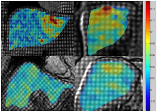
Liver disease causes millions of deaths per year worldwide, and approximately half of these cases are due to cirrhosis, which is an advanced stage of liver fibrosis that can be accompanied by liver failure and portal hypertension. Early detection of liver fibrosis helps in improving its treatment and prevents its progression to cirrhosis. In this work, we present a novel noninvasive method to detect liver fibrosis from tagged MRI images using a machine learning-based approach. Specifically, coronal and sagittal tagged MRI imaging are analyzed separately to capture cardiac-induced deformation of the liver. The liver is manually delineated and a novel image feature, namely, the histogram of the peak strain (HPS) value, is computed from the segmented liver region and is used to classify the liver as being either normal or fibrotic. Classification is achieved using a support vector machine algorithm. The in vivo study included 15 healthy volunteers (10 males; age range 30-45 years) and 22 patients (15 males; age range 25-50 years) with liver fibrosis verified and graded by transient elastography, and 10 patients only had a liver biopsy and were diagnosed with a score of F3-F4. The proposed method demonstrates the usefulness and efficiency of extracting the HPS features from the sagittal slices for patients with moderate fibrosis. Cross-validation of the method showed an accuracy of 83.7% (specificity = 86.6%, sensitivity = 81.8%).
中文翻译:

使用基于机器学习的方法检测肝脏纤维化,以量化标记的MR图像中心脏诱发的变形。
肝病每年在世界范围内造成数百万的死亡,其中大约一半是由于肝硬化,这是肝纤维化的晚期,可伴有肝衰竭和门静脉高压症。早期发现肝纤维化有助于改善治疗并防止其发展为肝硬化。在这项工作中,我们提出了一种新颖的非侵入性方法,使用基于机器学习的方法从标记的MRI图像中检测肝纤维化。具体来说,分别分析冠状动脉和矢状位MRI成像以捕获心脏引起的肝脏变形。手动描绘肝脏,并从分割的肝脏区域计算出新的图像特征,即峰值应变(HPS)值的直方图,并用于将肝脏分类为正常或纤维化。使用支持向量机算法可实现分类。体内研究包括15名健康志愿者(10名男性;年龄范围30-45岁)和22例患者(15名男性;年龄范围25-50岁),并通过瞬时弹性成像证实了肝纤维化并对其进行了分级,其中10例患者只有肝脏活检并被诊断为F3-F4评分。所提出的方法证明了从矢状切片中提取HPS特征对于中度纤维化患者的有用性和有效性。该方法的交叉验证显示准确度为83.7%(特异性= 86.6%,灵敏度= 81.8%)。10例仅进行了肝活检,并被诊断为F3-F4评分。所提出的方法证明了从矢状切片中提取HPS特征对于中度纤维化患者的有用性和有效性。该方法的交叉验证显示准确度为83.7%(特异性= 86.6%,灵敏度= 81.8%)。10例仅进行了肝活检,并被诊断为F3-F4评分。所提出的方法证明了从矢状切片中提取HPS特征对于中度纤维化患者的有用性和有效性。该方法的交叉验证显示准确度为83.7%(特异性= 86.6%,灵敏度= 81.8%)。
更新日期:2019-12-20
中文翻译:
使用基于机器学习的方法检测肝脏纤维化,以量化标记的MR图像中心脏诱发的变形。
肝病每年在世界范围内造成数百万的死亡,其中大约一半是由于肝硬化,这是肝纤维化的晚期,可伴有肝衰竭和门静脉高压症。早期发现肝纤维化有助于改善治疗并防止其发展为肝硬化。在这项工作中,我们提出了一种新颖的非侵入性方法,使用基于机器学习的方法从标记的MRI图像中检测肝纤维化。具体来说,分别分析冠状动脉和矢状位MRI成像以捕获心脏引起的肝脏变形。手动描绘肝脏,并从分割的肝脏区域计算出新的图像特征,即峰值应变(HPS)值的直方图,并用于将肝脏分类为正常或纤维化。使用支持向量机算法可实现分类。体内研究包括15名健康志愿者(10名男性;年龄范围30-45岁)和22例患者(15名男性;年龄范围25-50岁),并通过瞬时弹性成像证实了肝纤维化并对其进行了分级,其中10例患者只有肝脏活检并被诊断为F3-F4评分。所提出的方法证明了从矢状切片中提取HPS特征对于中度纤维化患者的有用性和有效性。该方法的交叉验证显示准确度为83.7%(特异性= 86.6%,灵敏度= 81.8%)。10例仅进行了肝活检,并被诊断为F3-F4评分。所提出的方法证明了从矢状切片中提取HPS特征对于中度纤维化患者的有用性和有效性。该方法的交叉验证显示准确度为83.7%(特异性= 86.6%,灵敏度= 81.8%)。10例仅进行了肝活检,并被诊断为F3-F4评分。所提出的方法证明了从矢状切片中提取HPS特征对于中度纤维化患者的有用性和有效性。该方法的交叉验证显示准确度为83.7%(特异性= 86.6%,灵敏度= 81.8%)。


 京公网安备 11010802027423号