当前位置:
X-MOL 学术
›
J. Biophotonics
›
论文详情
Our official English website, www.x-mol.net, welcomes your
feedback! (Note: you will need to create a separate account there.)
Full-field swept-source optical coherence tomography and neural tissue classification for deep brain imaging.
Journal of Biophotonics ( IF 2.0 ) Pub Date : 2019-12-02 , DOI: 10.1002/jbio.201960083 Ilan Felts Almog 1, 2 , Fu-Der Chen 1, 2 , Suhan Senova 2, 3, 4 , Anton Fomenko 2 , Elise Gondard 2 , Wesley D Sacher 1, 5 , Andres M Lozano 2, 6 , Joyce K S Poon 1, 2, 5
Journal of Biophotonics ( IF 2.0 ) Pub Date : 2019-12-02 , DOI: 10.1002/jbio.201960083 Ilan Felts Almog 1, 2 , Fu-Der Chen 1, 2 , Suhan Senova 2, 3, 4 , Anton Fomenko 2 , Elise Gondard 2 , Wesley D Sacher 1, 5 , Andres M Lozano 2, 6 , Joyce K S Poon 1, 2, 5
Affiliation
|
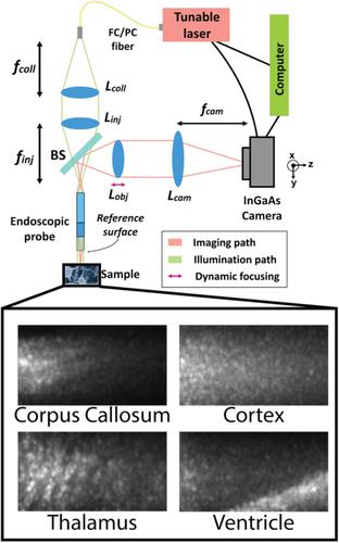
Optical coherence tomography can differentiate brain regions with intrinsic contrast and at a micron scale resolution. Such a device can be particularly useful as a real‐time neurosurgical guidance tool. We present, to our knowledge, the first full‐field swept‐source optical coherence tomography system operating near a wavelength of 1310 nm. The proof‐of‐concept system was integrated with an endoscopic probe tip, which is compatible with deep brain stimulation keyhole neurosurgery. Neuroimaging experiments were performed on ex vivo brain tissues and in vivo in rat brains. Using classification algorithms involving texture features and optical attenuation, images were successfully classified into three brain tissue types.
中文翻译:

用于深部脑成像的全场扫源光学相干断层扫描和神经组织分类。
光学相干断层扫描可以通过内在对比度和微米级分辨率来区分大脑区域。这种设备作为实时神经外科指导工具特别有用。据我们所知,我们推出了第一个全场扫频光学相干断层扫描系统,工作波长接近 1310 nm。该概念验证系统与内窥镜探头尖端集成,与深部脑刺激锁孔神经外科手术兼容。在离体脑组织和体内大鼠脑中进行神经影像学实验。使用涉及纹理特征和光学衰减的分类算法,图像被成功分类为三种脑组织类型。
更新日期:2019-12-02
中文翻译:
用于深部脑成像的全场扫源光学相干断层扫描和神经组织分类。
光学相干断层扫描可以通过内在对比度和微米级分辨率来区分大脑区域。这种设备作为实时神经外科指导工具特别有用。据我们所知,我们推出了第一个全场扫频光学相干断层扫描系统,工作波长接近 1310 nm。该概念验证系统与内窥镜探头尖端集成,与深部脑刺激锁孔神经外科手术兼容。在离体脑组织和体内大鼠脑中进行神经影像学实验。使用涉及纹理特征和光学衰减的分类算法,图像被成功分类为三种脑组织类型。


 京公网安备 11010802027423号