Magnetic Resonance Imaging ( IF 2.1 ) Pub Date : 2019-11-11 , DOI: 10.1016/j.mri.2019.11.002 Mehran Azimbagirad , Fabrício H. Simozo , Antonio C.S. Senra Filho , Luiz O. Murta Junior
|
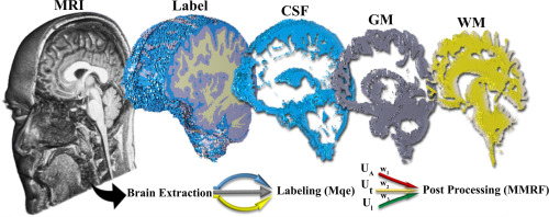
Quantifying the intracranial tissue volume changes in magnetic resonance imaging (MRI) assists specialists to analyze the effects of natural or pathological changes. Since these changes can be subtle, the accuracy of the automatic compartmentalization method is always criticized by specialists. We propose and then evaluate an automatic segmentation method based on modified q-entropy (Mqe) through a modified Markov Random Field (MMRF) enhanced by Alzheimer anatomic reference (AAR) to provide a high accuracy brain tissues parcellation approach (Mqe-MMRF). We underwent two strategies to evaluate Mqe-MMRF; a simulation of different levels of noise and non-uniformity effect on MRI data (7 subjects) and a set of twenty MRI data available from MRBrainS13 as patient brain tissue segmentation challenge. We accessed eleven quality metrics compared to reference tissues delineations to evaluate Mqe-MMRF. MRI segmentation scores decreased by only 4.6% on quality metrics after noise and non-uniformity simulations of 40% and 9%, respectively. We found significant mean improvements in the metrics of the five training subjects, for whole-brain 0.86%, White Matter 3.20%, Gray Matter 3.99%, and Cerebrospinal Fluid 4.16% (p-values < 0.02) when Mqe-MMRF compared to the other reference methods. We also processed the Mqe-MMRF on 15 evaluation subjects group from MRBrainS13 online challenge, and the results held a higher rank than the reference tools; FreeSurfer, SPM, and FSL. Since the proposed method improved the precision of brain segmentation, specifically, for GM, and thus one can use it in quantitative and morphological brain studies.
中文翻译:
通过MRF和Alzheimer解剖参考进行Tsallis熵分割,以进行脑部磁共振消融
在磁共振成像(MRI)中量化颅内组织体积变化有助于专家分析自然或病理变化的影响。由于这些更改可能是微妙的,因此自动分隔方法的准确性始终受到专家的批评。我们提出然后评估通过基于改进的马尔可夫随机场(MMRF)和改进的q熵(Mqe)的自动分割方法,该改进的马尔科夫随机场由阿尔茨海默病解剖参考(AAR)增强,以提供高精度的脑组织切碎方法(Mqe-MMRF)。我们采用了两种策略来评估Mqe-MMRF;对MRI数据(7名受试者)的不同水平的噪声和非均匀性影响进行了模拟,并从MRBrainS13获得了20份MRI数据作为患者脑组织分割挑战。我们访问了11个质量指标,与参考组织的轮廓进行了比较,以评估Mqe-MMRF。在分别进行40%和9%的噪声和非均匀性模拟后,按质量度量标准的MRI分割分数仅下降了4.6%。我们发现,与Mqe-MMRF相比,五项训练对象的指标的全脑平均改善显着,全脑0.86%,白质3.20%,灰质3.99%和脑脊液4.16%(p值<0.02)。其他参考方法。我们还处理了来自MRBrainS13在线挑战赛的15个评估主题组的Mqe-MMRF,其结果比参考工具具有更高的排名;FreeSurfer,SPM和FSL。由于所提出的方法提高了脑部分割的精确度,特别是针对GM,因此可以将其用于定量和形态学脑部研究。


 京公网安备 11010802027423号