当前位置:
X-MOL 学术
›
Surf. Sci.
›
论文详情
Our official English website, www.x-mol.net, welcomes your
feedback! (Note: you will need to create a separate account there.)
Pristine and oxidised Ag-nanoparticles on free-standing graphene as explored by X-ray photoelectron and Auger spectroscopy
Surface Science ( IF 2.1 ) Pub Date : 2020-03-01 , DOI: 10.1016/j.susc.2019.121533 M. Al-Hada , L. Gregoratti , M. Amati , M. Neeb
Surface Science ( IF 2.1 ) Pub Date : 2020-03-01 , DOI: 10.1016/j.susc.2019.121533 M. Al-Hada , L. Gregoratti , M. Amati , M. Neeb
|
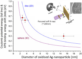
Abstract Silver nanoparticles with a mean diameter of 15 nm have been grown on free-standing graphene and explored by soft X-ray synchrotron radiation using the ESCAmicroscopy beamline at Elettra. In particular, the 3d core orbitals and valence band (4d/5 s) of pristine and plasma-oxidised Ag-nanoparticles have been investigated by X-ray photoelectron spectroscopy (XPS), X-ray induced valence band photoemission, and MNN Auger spectroscopy. The X-ray spectra of the pristine Ag-nanoparticles, i.e. XPS, Auger and UPS spectra, are very similar to these of the metallic Ag-bulk film while the spectra of the oxidised Ag-nanoparticles exhibit distinct differences with respect to the oxidised Ag-bulk film. This is inferred from a larger valence band width, an extended valence band photoemission intensity nearly up to the Fermi level of the bulk metal, a smaller two-hole Coulomb correlation energy and a rather bulk-like Auger spectrum. We interpret this as to result from a shell-like composition of the Ag-nanoparticles with a thin passivating oxide layer and an inner metallic core. Moreover, the 3d photoemission peak of the oxidised nanoparticles is shifted to higher binding energy with respect to the bulk oxide peak. This is attributed not only to an XPS chemical shift but also to an additional final state Coulomb charging according to the electrostatic liquid drop model. Comparing the core binding energy shift of Ag-nanoparticles with the electrostatic potential of a spherical compact particle and a flat nanoislands a morphology transformation of the oxidised nanoparticles from 3D to 2D is obvious in the size range 5–15 nm.
中文翻译:

通过 X 射线光电子和俄歇光谱探索的独立石墨烯上的原始和氧化银纳米颗粒
摘要 平均直径为 15 nm 的银纳米粒子已在独立的石墨烯上生长,并使用 Elettra 的 ESCA 显微镜光束线通过软 X 射线同步辐射进行探索。特别是,通过 X 射线光电子能谱 (XPS)、X 射线诱导价带光电发射和 MNN 俄歇光谱研究了原始和等离子体氧化的银纳米颗粒的 3d 核心轨道和价带 (4d/5 s) . 原始 Ag 纳米粒子的 X 射线光谱,即 XPS、俄歇和 UPS 光谱,与金属 Ag 体膜的 X 射线光谱非常相似,而氧化的 Ag 纳米粒子的光谱相对于氧化的 Ag 表现出明显的差异-散装电影。这是从更大的价带宽度推断出来的,扩展的价带光电发射强度几乎高达块状金属的费米能级,较小的两孔库仑相关能量和相当大的俄歇谱。我们将此解释为由具有薄钝化氧化物层和内部金属核的 Ag 纳米颗粒的壳状组合物引起的。此外,氧化纳米粒子的 3d 光电发射峰相对于体氧化物峰转移到更高的结合能。这不仅归因于 XPS 化学位移,而且归因于根据静电液滴模型的额外终态库仑充电。将 Ag 纳米颗粒的核心结合能位移与球形致密颗粒和扁平纳米岛的静电势进行比较,氧化纳米颗粒从 3D 到 2D 的形态转变在 5-15 nm 的尺寸范围内是显而易见的。
更新日期:2020-03-01
中文翻译:
通过 X 射线光电子和俄歇光谱探索的独立石墨烯上的原始和氧化银纳米颗粒
摘要 平均直径为 15 nm 的银纳米粒子已在独立的石墨烯上生长,并使用 Elettra 的 ESCA 显微镜光束线通过软 X 射线同步辐射进行探索。特别是,通过 X 射线光电子能谱 (XPS)、X 射线诱导价带光电发射和 MNN 俄歇光谱研究了原始和等离子体氧化的银纳米颗粒的 3d 核心轨道和价带 (4d/5 s) . 原始 Ag 纳米粒子的 X 射线光谱,即 XPS、俄歇和 UPS 光谱,与金属 Ag 体膜的 X 射线光谱非常相似,而氧化的 Ag 纳米粒子的光谱相对于氧化的 Ag 表现出明显的差异-散装电影。这是从更大的价带宽度推断出来的,扩展的价带光电发射强度几乎高达块状金属的费米能级,较小的两孔库仑相关能量和相当大的俄歇谱。我们将此解释为由具有薄钝化氧化物层和内部金属核的 Ag 纳米颗粒的壳状组合物引起的。此外,氧化纳米粒子的 3d 光电发射峰相对于体氧化物峰转移到更高的结合能。这不仅归因于 XPS 化学位移,而且归因于根据静电液滴模型的额外终态库仑充电。将 Ag 纳米颗粒的核心结合能位移与球形致密颗粒和扁平纳米岛的静电势进行比较,氧化纳米颗粒从 3D 到 2D 的形态转变在 5-15 nm 的尺寸范围内是显而易见的。


 京公网安备 11010802027423号