Modern Pathology ( IF 7.1 ) Pub Date : 2019-11-07 , DOI: 10.1038/s41379-019-0409-3 Seung-Mo Hong 1, 2 , DongJun Jung 1, 3 , Ashley Kiemen 4 , Matthias M Gaida 5, 6 , Tadashi Yoshizawa 1 , Alicia M Braxton 1 , Michaël Noë 7 , Gemma Lionheart 1 , Kiyoko Oshima 1 , Elizabeth D Thompson 1, 7 , Richard Burkhart 8 , Pei-Hsun Wu 4 , Denis Wirtz 1, 4 , Ralph H Hruban 1, 7 , Laura D Wood 1, 7
|
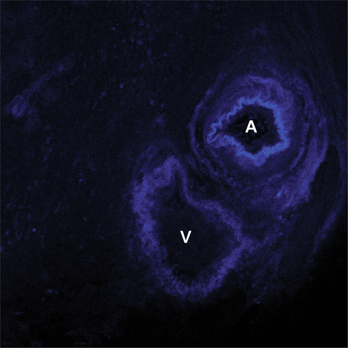
Venous invasion is three times more common in pancreatic cancer than it is in other major cancers of the gastrointestinal tract, and venous invasion may explain why pancreatic cancer is so deadly. To characterize the patterns of venous invasion in pancreatic cancer, 52 thick slabs (up to 5 mm) of tissue were harvested from 52 surgically resected human ductal adenocarcinomas, cleared with a modified iDISCO method, and labeled with fluorescent-conjugated antibodies to cytokeratin 19, desmin, CD31, p53 and/or e-cadherin. Labeled three-dimensional (3D) pancreas cancer tissues were visualized with confocal laser scanning or light sheet microscopy. Multiple foci of venous and even arterial invasion were visualized. Venous invasion was detected more often in 3D (88%, 30/34 cases) than in conventional 2D slide evaluation (75%, 25/34 cases, P < 0.001). 3D visualization revealed pancreatic cancer cells crossing the walls of veins at multiple points, often at points where preexisting capillary structures bridge the blood vessels. The neoplastic cells often retained a ductal morphology (cohesive cells forming tubes) as they progressed from a stromal to intravenous location. Although immunolabeling with antibodies to e-cadherin revealed focal loss of expression at the leading edges of the cancers, the neoplastic cells within veins expressed e-cadherin and formed well-oriented glands. We conclude that venous invasion is almost universal in pancreatic cancer, suggesting that even surgically resectable PDAC has access to the venous spaces and thus the ability to disseminate widely. Furthermore, we observe that sustained epithelial–mesenchymal transition is not required for venous invasion in pancreatic cancer.
中文翻译:
清除的人胰腺癌的三维可视化显示,静脉侵入不需要持续的上皮-间质转化。
胰腺癌中静脉侵犯的发生率是其他主要胃肠道癌症的三倍,静脉侵犯可以解释为什么胰腺癌如此致命。为了表征胰腺癌静脉侵犯的模式,从 52 例手术切除的人导管腺癌中采集了 52 块厚板(最多 5 毫米)的组织,用改良的 iDISCO 方法进行清除,并用细胞角蛋白 19 的荧光偶联抗体进行标记,结蛋白、CD31、p53 和/或 e-钙粘蛋白。使用共焦激光扫描或光片显微镜观察标记的三维 (3D) 胰腺癌组织。可见多个静脉甚至动脉侵犯灶。与传统的 2D 玻片评估(75%,25/34 例, P < 0.001)相比,3D 中更常检测到静脉侵犯(88%,30/34 例) 。3D 可视化显示胰腺癌细胞在多个点穿过静脉壁,通常是在先前存在的毛细血管结构桥接血管的点。当肿瘤细胞从基质位置进展到静脉位置时,它们通常保留导管形态(形成管的粘性细胞)。尽管e-钙粘蛋白抗体的免疫标记显示癌症前缘局部表达缺失,但静脉内的肿瘤细胞表达e-钙粘蛋白并形成定向良好的腺体。我们得出的结论是,静脉侵犯在胰腺癌中几乎是普遍存在的,这表明即使是可手术切除的 PDAC 也可以进入静脉空间,从而能够广泛传播。此外,我们观察到胰腺癌的静脉侵犯不需要持续的上皮-间质转化。


 京公网安备 11010802027423号