当前位置:
X-MOL 学术
›
Modern Pathol.
›
论文详情
Our official English website, www.x-mol.net, welcomes your feedback! (Note: you will need to create a separate account there.)
Cyclin D1 immunohistochemical staining to separate benign from malignant mesothelial proliferations.
Modern Pathology ( IF 7.5 ) Pub Date : 2019-11-04 , DOI: 10.1038/s41379-019-0411-9 Jennifer Pors 1 , Julia Naso 1 , Kyra Berg 1 , Andrew Churg 1
Modern Pathology ( IF 7.5 ) Pub Date : 2019-11-04 , DOI: 10.1038/s41379-019-0411-9 Jennifer Pors 1 , Julia Naso 1 , Kyra Berg 1 , Andrew Churg 1
Affiliation
|
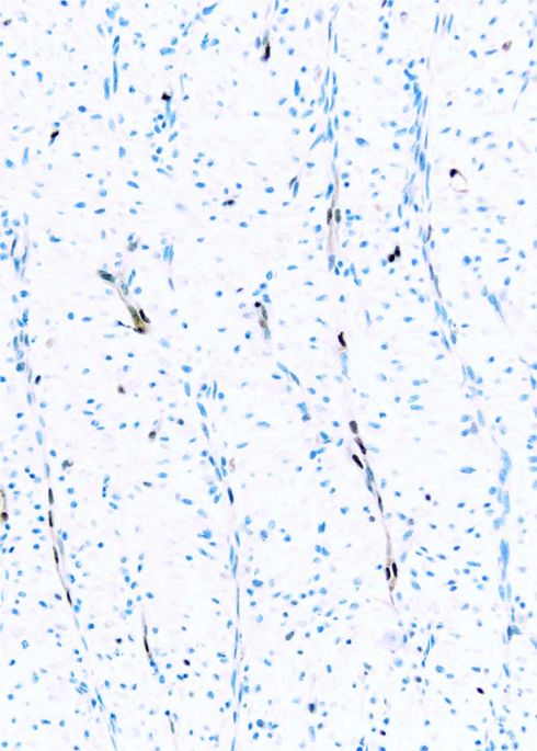
The separation of benign from malignant mesothelial proliferations is a morphologically difficult problem. Mutations/deletions of components of the Hippo pathway are frequent in malignant mesotheliomas, and one downstream effect of aberrant Hippo signaling is increased production of cyclin D1. We examined expression of cyclin D1 nuclear staining in two tissue microarrays containing 52 reactive epithelial mesothelial proliferations, 51 reactive spindle cell mesothelial proliferations, 54 epithelial mesotheliomas, and 22 sarcomatous/desmoplastic mesotheliomas. When present, cyclin D1 staining was always strong, hence the arrays were scored as 0, 1-25%, 26-50%, 51-75%, and 76-100% staining. Both arrays showed a similar pattern. Reactive epithelial proliferations generally showed no staining (42/52 cases) or 1-25% staining (10/52 cases) with no cases showing >25% staining. Overall for reactive epithelial proliferations the maximum staining was 14.8% and mean 1.1 ± 2.9%. For epithelial mesotheliomas 39/54 (72%) cases demonstrated >25% staining, with 8/54 in the 26-50% staining range, 9/54 in the 51-75% range, and 22/54 in the >75% range. Combinations of staining using cyclin D1 >50% plus BAP1 or MTAP loss in epithelial mesotheliomas produced about a 10% increase in sensitivity. Reactive spindle cell proliferations showed a broader range of staining with 27/51 in the 1-25% range, 5/51 in the 26-50% range, and 1/51 >50%. Eleven of 22 sarcomatous/desmoplastic mesotheliomas scored 50% or greater. We conclude that for epithelial mesothelial proliferations, the finding of >50% of tumor cells staining supports a diagnosis of epithelial mesothelioma with 100% specificity but only modest (57%) sensitivity.
中文翻译:

细胞周期蛋白 D1 免疫组织化学染色可区分良性和恶性间皮增生。
从形态学上区分良性和恶性间皮增生是一个难题。Hippo 通路成分的突变/缺失在恶性间皮瘤中很常见,异常的 Hippo 信号通路的一个下游效应是细胞周期蛋白 D1 的产生增加。我们检查了两个组织微阵列中细胞周期蛋白 D1 核染色的表达,其中包含 52 个反应性上皮间皮增生、51 个反应性梭形细胞间皮增生、54 个上皮间皮瘤和 22 个肉瘤/促纤维增生性间皮瘤。当存在时,细胞周期蛋白 D1 染色始终很强,因此阵列评分为 0、1-25%、26-50%、51-75% 和 76-100% 染色。两个阵列都显示出相似的模式。反应性上皮增生通常显示无染色(42/52 例)或 1-25% 染色(10/52 例),没有显示>25% 染色的病例。总的来说,反应性上皮增生的最大染色为 14.8%,平均为 1.1 ± 2.9%。对于上皮间皮瘤,39/54 (72%) 例显示 >25% 染色,其中 8/54 在 26-50% 染色范围内,9/54 在 51-75% 范围内,22/54 在 >75% 范围内范围。在上皮间皮瘤中使用细胞周期蛋白 D1 >50% 加上 BAP1 或 MTAP 损失的染色组合产生了大约 10% 的灵敏度增加。反应性梭形细胞增殖显示更广泛的染色范围,27/51 在 1-25% 范围内,5/51 在 26-50% 范围内,1/51 >50%。22 例肉瘤/促纤维增生性间皮瘤中有 11 例评分为 50% 或更高。我们得出结论,对于上皮间皮增生,
更新日期:2019-11-04
中文翻译:
细胞周期蛋白 D1 免疫组织化学染色可区分良性和恶性间皮增生。
从形态学上区分良性和恶性间皮增生是一个难题。Hippo 通路成分的突变/缺失在恶性间皮瘤中很常见,异常的 Hippo 信号通路的一个下游效应是细胞周期蛋白 D1 的产生增加。我们检查了两个组织微阵列中细胞周期蛋白 D1 核染色的表达,其中包含 52 个反应性上皮间皮增生、51 个反应性梭形细胞间皮增生、54 个上皮间皮瘤和 22 个肉瘤/促纤维增生性间皮瘤。当存在时,细胞周期蛋白 D1 染色始终很强,因此阵列评分为 0、1-25%、26-50%、51-75% 和 76-100% 染色。两个阵列都显示出相似的模式。反应性上皮增生通常显示无染色(42/52 例)或 1-25% 染色(10/52 例),没有显示>25% 染色的病例。总的来说,反应性上皮增生的最大染色为 14.8%,平均为 1.1 ± 2.9%。对于上皮间皮瘤,39/54 (72%) 例显示 >25% 染色,其中 8/54 在 26-50% 染色范围内,9/54 在 51-75% 范围内,22/54 在 >75% 范围内范围。在上皮间皮瘤中使用细胞周期蛋白 D1 >50% 加上 BAP1 或 MTAP 损失的染色组合产生了大约 10% 的灵敏度增加。反应性梭形细胞增殖显示更广泛的染色范围,27/51 在 1-25% 范围内,5/51 在 26-50% 范围内,1/51 >50%。22 例肉瘤/促纤维增生性间皮瘤中有 11 例评分为 50% 或更高。我们得出结论,对于上皮间皮增生,


 京公网安备 11010802027423号