当前位置:
X-MOL 学术
›
Cytom. Part A
›
论文详情
Our official English website, www.x-mol.net, welcomes your
feedback! (Note: you will need to create a separate account there.)
Collagen Organization Within the Cartilage of Trpv4-/- Mice Studied with Two-Photon Microscopy and Polarized Second Harmonic Generation.
Cytometry Part A ( IF 2.5 ) Pub Date : 2019-10-11 , DOI: 10.1002/cyto.a.23900 M Rocio Servin-Vences 1 , Kate Poole 1 , Anje Sporbert 2 , Gary R Lewin 1 , Anca Margineanu 2
Cytometry Part A ( IF 2.5 ) Pub Date : 2019-10-11 , DOI: 10.1002/cyto.a.23900 M Rocio Servin-Vences 1 , Kate Poole 1 , Anje Sporbert 2 , Gary R Lewin 1 , Anca Margineanu 2
Affiliation
|
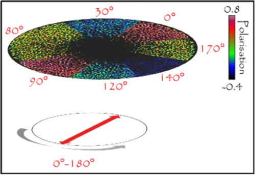
The polymodal channel TRPV4 has been shown to regulate development and maintenance of cartilage. Here we investigate whether TRPV4 activity regulates the early deposition and structure of collagen matrix in the femoral head cartilage by comparing the 3D morphology and the sub-micrometer organization of the collagen matrix between wild type and Trpv4 -/- mice pups four to five days old. Two-photon microscopy can be used to conduct label-free imaging of cartilage, as collagen generates a second harmonic signal (second harmonic generation [SHG]) under pulsed infrared excitation. In one set of measurements, we use circularly polarized laser light to reconstruct the 3D morphology of the femoral head cartilage and to measure the tissue thickness. Second, by rotating the direction of the linearly polarized light and using polarized SHG detection, we investigate the sub-micrometer orientation of collagen fibers in the cartilage. At this developmental stage, we cannot detect statistically significant differences between the two mice strains, although a tendency toward a more random orientation of collagen fibers and a higher thickness of the whole cartilage seems to characterize the Trpv4 -/- mice. We discuss possible reasons for these observations. © 2019 The Authors. Cytometry Part A published by Wiley Periodicals, Inc. on behalf of International Society for Advancement of Cytometry.
中文翻译:

用双光子显微镜和偏振二次谐波产生研究 Trpv4-/- 小鼠软骨内的胶原组织。
多模式通道 TRPV4 已被证明可以调节软骨的发育和维持。在这里,我们通过比较野生型和 Trpv4 -/- 小鼠幼崽之间的 3D 形态和胶原基质的亚微米组织来研究 TRPV4 活性是否调节股骨头软骨中胶原基质的早期沉积和结构. 双光子显微镜可用于对软骨进行无标记成像,因为胶原蛋白在脉冲红外激发下会产生二次谐波信号(二次谐波生成 [SHG])。在一组测量中,我们使用圆偏振激光重建股骨头软骨的 3D 形态并测量组织厚度。其次,通过旋转线偏振光的方向并使用偏振SHG检测,我们研究了软骨中胶原纤维的亚微米方向。在这个发育阶段,我们无法检测到两种小鼠品系之间的统计学显着差异,尽管 Trpv4 -/- 小鼠的特征似乎是胶原纤维更随机的方向和整个软骨的厚度更高。我们讨论了这些观察结果的可能原因。© 2019 作者。Cytometry Part A 由 Wiley Periodicals, Inc. 代表 International Society for Advancement of Cytometry 出版。尽管倾向于更随机的胶原纤维取向和更高厚度的整个软骨似乎是 Trpv4 -/- 小鼠的特征。我们讨论了这些观察结果的可能原因。© 2019 作者。Cytometry Part A 由 Wiley Periodicals, Inc. 代表 International Society for Advancement of Cytometry 出版。尽管倾向于更随机的胶原纤维取向和更高厚度的整个软骨似乎是 Trpv4 -/- 小鼠的特征。我们讨论了这些观察结果的可能原因。© 2019 作者。Cytometry Part A 由 Wiley Periodicals, Inc. 代表 International Society for Advancement of Cytometry 出版。
更新日期:2019-10-11
中文翻译:
用双光子显微镜和偏振二次谐波产生研究 Trpv4-/- 小鼠软骨内的胶原组织。
多模式通道 TRPV4 已被证明可以调节软骨的发育和维持。在这里,我们通过比较野生型和 Trpv4 -/- 小鼠幼崽之间的 3D 形态和胶原基质的亚微米组织来研究 TRPV4 活性是否调节股骨头软骨中胶原基质的早期沉积和结构. 双光子显微镜可用于对软骨进行无标记成像,因为胶原蛋白在脉冲红外激发下会产生二次谐波信号(二次谐波生成 [SHG])。在一组测量中,我们使用圆偏振激光重建股骨头软骨的 3D 形态并测量组织厚度。其次,通过旋转线偏振光的方向并使用偏振SHG检测,我们研究了软骨中胶原纤维的亚微米方向。在这个发育阶段,我们无法检测到两种小鼠品系之间的统计学显着差异,尽管 Trpv4 -/- 小鼠的特征似乎是胶原纤维更随机的方向和整个软骨的厚度更高。我们讨论了这些观察结果的可能原因。© 2019 作者。Cytometry Part A 由 Wiley Periodicals, Inc. 代表 International Society for Advancement of Cytometry 出版。尽管倾向于更随机的胶原纤维取向和更高厚度的整个软骨似乎是 Trpv4 -/- 小鼠的特征。我们讨论了这些观察结果的可能原因。© 2019 作者。Cytometry Part A 由 Wiley Periodicals, Inc. 代表 International Society for Advancement of Cytometry 出版。尽管倾向于更随机的胶原纤维取向和更高厚度的整个软骨似乎是 Trpv4 -/- 小鼠的特征。我们讨论了这些观察结果的可能原因。© 2019 作者。Cytometry Part A 由 Wiley Periodicals, Inc. 代表 International Society for Advancement of Cytometry 出版。


 京公网安备 11010802027423号