当前位置:
X-MOL 学术
›
Cytom. Part A
›
论文详情
Our official English website, www.x-mol.net, welcomes your
feedback! (Note: you will need to create a separate account there.)
Estimation of the Mitochondrial Membrane Potential Using Fluorescence Lifetime Imaging Microscopy.
Cytometry Part A ( IF 2.5 ) Pub Date : 2019-09-05 , DOI: 10.1002/cyto.a.23886 Irina A Okkelman 1 , Dmitri B Papkovsky 1 , Ruslan I Dmitriev 1, 2
Cytometry Part A ( IF 2.5 ) Pub Date : 2019-09-05 , DOI: 10.1002/cyto.a.23886 Irina A Okkelman 1 , Dmitri B Papkovsky 1 , Ruslan I Dmitriev 1, 2
Affiliation
|
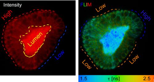
Monitoring of cell metabolism represents an important application area for fluorescence lifetime imaging microscopy (FLIM). In particular, assessment of mitochondrial membrane potential (MMP) in complex three-dimensional multicellular in vitro, ex vivo, and in vivo models would enable improved segmentation and functional discrimination of cell types, directly report on the mitochondrial function and complement the quenched-phosphorescence detection of cellular O2 and two-photon excited FLIM of endogenous NAD(P)H. Here, we report the green and orange-emitting fluorescent dyes SYTO and tetramethylrhodamine methyl ester (TMRM) as potential FLIM probes for MMP. In addition to nuclear, SYTO 16 and 24 dyes also display mitochondrial accumulation. FLIM with the culture of human colon cancer HCT116 cells allowed observation of the heterogeneity of mitochondrial polarization during the cell cycle progression. The dyes also demonstrated good performance with 3D cultures of Lgr5-GFP mouse intestinal organoids, providing efficient and quick cell staining and compatibility with two-photon excitation. Multiplexed imaging of Lgr5-GFP, proliferating cells (Hoechst 33342-aided FLIM), and TMRM-FLIM allowed us to identify the population of metabolically active cells in stem cell niche. TMRM-FLIM enabled to visualize the differences in membrane potential between Lgr5-positive and other proliferating and differentiated cell types. Altogether, SYTO 24 and TMRM dyes represent promising markers for advanced FLIM-based studies of cell bioenergetics with complex 3D and in vivo models. © 2019 International Society for Advancement of Cytometry.
中文翻译:

使用荧光寿命成像显微镜估计线粒体膜电位。
细胞代谢监测代表了荧光寿命成像显微镜 (FLIM) 的一个重要应用领域。特别是,在体外、离体和体内复杂的三维多细胞模型中评估线粒体膜电位 (MMP) 将能够改进细胞类型的分割和功能区分,直接报告线粒体功能并补充淬灭的磷光检测细胞 O2 和双光子激发内源性 NAD(P)H 的 FLIM。在这里,我们报告了绿色和橙色荧光染料 SYTO 和四甲基罗丹明甲酯 (TMRM) 作为 MMP 的潜在 FLIM 探针。除了核外,SYTO 16 和 24 染料也显示出线粒体积累。FLIM 与人结肠癌 HCT116 细胞培养物允许观察细胞周期进程中线粒体极化的异质性。这些染料在 Lgr5-GFP 小鼠肠道类器官的 3D 培养中也表现出良好的性能,提供高效、快速的细胞染色以及与双光子激发的兼容性。Lgr5-GFP、增殖细胞(Hoechst 33342 辅助 FLIM)和 TMRM-FLIM 的多重成像使我们能够识别干细胞生态位中代谢活跃的细胞群。TMRM-FLIM 能够可视化 Lgr5 阳性和其他增殖和分化细胞类型之间的膜电位差异。总之,SYTO 24 和 TMRM 染料代表了具有复杂 3D 和体内模型的基于 FLIM 的高级细胞生物能量学研究的有希望的标记。
更新日期:2019-09-05
中文翻译:
使用荧光寿命成像显微镜估计线粒体膜电位。
细胞代谢监测代表了荧光寿命成像显微镜 (FLIM) 的一个重要应用领域。特别是,在体外、离体和体内复杂的三维多细胞模型中评估线粒体膜电位 (MMP) 将能够改进细胞类型的分割和功能区分,直接报告线粒体功能并补充淬灭的磷光检测细胞 O2 和双光子激发内源性 NAD(P)H 的 FLIM。在这里,我们报告了绿色和橙色荧光染料 SYTO 和四甲基罗丹明甲酯 (TMRM) 作为 MMP 的潜在 FLIM 探针。除了核外,SYTO 16 和 24 染料也显示出线粒体积累。FLIM 与人结肠癌 HCT116 细胞培养物允许观察细胞周期进程中线粒体极化的异质性。这些染料在 Lgr5-GFP 小鼠肠道类器官的 3D 培养中也表现出良好的性能,提供高效、快速的细胞染色以及与双光子激发的兼容性。Lgr5-GFP、增殖细胞(Hoechst 33342 辅助 FLIM)和 TMRM-FLIM 的多重成像使我们能够识别干细胞生态位中代谢活跃的细胞群。TMRM-FLIM 能够可视化 Lgr5 阳性和其他增殖和分化细胞类型之间的膜电位差异。总之,SYTO 24 和 TMRM 染料代表了具有复杂 3D 和体内模型的基于 FLIM 的高级细胞生物能量学研究的有希望的标记。


 京公网安备 11010802027423号