当前位置:
X-MOL 学术
›
Cytom. Part A
›
论文详情
Our official English website, www.x-mol.net, welcomes your
feedback! (Note: you will need to create a separate account there.)
Diffusion Mapping of Eosinophil-Activation State.
Cytometry Part A ( IF 2.5 ) Pub Date : 2019-08-31 , DOI: 10.1002/cyto.a.23884 Justyna Piasecka 1 , Catherine A Thornton 1 , Paul Rees 2 , Huw D Summers 2
Cytometry Part A ( IF 2.5 ) Pub Date : 2019-08-31 , DOI: 10.1002/cyto.a.23884 Justyna Piasecka 1 , Catherine A Thornton 1 , Paul Rees 2 , Huw D Summers 2
Affiliation
|
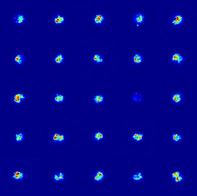
Eosinophils are granular leukocytes that play a role in mediating inflammatory responses linked to infection and allergic disease. Their activation during an immune response triggers spatial reorganization and eventual cargo release from intracellular granules. Understanding this process is important in diagnosing eosinophilic disorders and in assessing treatment efficacy; however, current protocols are limited to simply quantifying the number of eosinophils within a blood sample. Given that high optical absorption and scattering by the granular structure of these cells lead to marked image features, the physical changes that occur during activation should be trackable using image analysis. Here, we present a study in which imaging flow cytometry is used to quantify eosinophil activation state, based on the extraction of 85 distinct spatial features from dark-field images formed by light scattered orthogonally to the illuminating beam. We apply diffusion mapping, a time inference method that orders cells on a trajectory based on similar image features. Analysis of exogenous cell activation using eotaxin and endogenous activation in donor samples with elevated eosinophil counts shows that cell position along the diffusion-path line correlates with activation level (99% confidence level). Thus, the diffusion mapping provides an activation metric for each cell. Assessment of activated and control populations using both this spatial image-based, activation score and the integrated side-scatter intensity shows an improved Fisher discriminant ratio rd = 0.7 for the multivariate technique compared with an rd = 0.47 for the traditional whole-cell scatter metric. © 2019 The Authors. Cytometry Part A published by Wiley Periodicals, Inc. on behalf of International Society for Advancement of Cytometry.
中文翻译:

嗜酸性粒细胞激活状态的扩散图。
嗜酸性粒细胞是颗粒白细胞,在介导与感染和过敏性疾病相关的炎症反应中发挥作用。它们在免疫反应期间的激活触发空间重组并最终从细胞内颗粒中释放货物。了解这一过程对于诊断嗜酸性粒细胞疾病和评估治疗效果非常重要;然而,目前的方案仅限于简单地量化血液样本中嗜酸性粒细胞的数量。鉴于这些细胞的颗粒结构的高光学吸收和散射会导致显着的图像特征,因此激活过程中发生的物理变化应该可以使用图像分析进行跟踪。在这里,我们提出了一项研究,其中成像流式细胞术用于量化嗜酸性粒细胞激活状态,基于从与照明光束正交散射的光形成的暗场图像中提取 85 个不同的空间特征。我们应用扩散映射,这是一种时间推断方法,可根据相似的图像特征对轨迹上的细胞进行排序。使用嗜酸性粒细胞计数升高的供体样品中使用嗜酸性粒细胞趋化因子进行的外源性细胞活化和内源性活化的分析表明,沿着扩散路径线的细胞位置与活化水平相关(99%置信水平)。因此,扩散映射为每个单元提供了激活度量。使用基于空间图像的激活分数和集成侧向散射强度对激活和对照群体进行评估,结果表明,与传统全细胞散射度量的 rd = 0.47 相比,多变量技术的 Fisher 判别比 rd = 0.7 得到了改进。© 2019 作者。细胞计数法 A 部分由 Wiley periodicals, Inc. 代表国际细胞计数法促进会出版。
更新日期:2020-03-09
中文翻译:
嗜酸性粒细胞激活状态的扩散图。
嗜酸性粒细胞是颗粒白细胞,在介导与感染和过敏性疾病相关的炎症反应中发挥作用。它们在免疫反应期间的激活触发空间重组并最终从细胞内颗粒中释放货物。了解这一过程对于诊断嗜酸性粒细胞疾病和评估治疗效果非常重要;然而,目前的方案仅限于简单地量化血液样本中嗜酸性粒细胞的数量。鉴于这些细胞的颗粒结构的高光学吸收和散射会导致显着的图像特征,因此激活过程中发生的物理变化应该可以使用图像分析进行跟踪。在这里,我们提出了一项研究,其中成像流式细胞术用于量化嗜酸性粒细胞激活状态,基于从与照明光束正交散射的光形成的暗场图像中提取 85 个不同的空间特征。我们应用扩散映射,这是一种时间推断方法,可根据相似的图像特征对轨迹上的细胞进行排序。使用嗜酸性粒细胞计数升高的供体样品中使用嗜酸性粒细胞趋化因子进行的外源性细胞活化和内源性活化的分析表明,沿着扩散路径线的细胞位置与活化水平相关(99%置信水平)。因此,扩散映射为每个单元提供了激活度量。使用基于空间图像的激活分数和集成侧向散射强度对激活和对照群体进行评估,结果表明,与传统全细胞散射度量的 rd = 0.47 相比,多变量技术的 Fisher 判别比 rd = 0.7 得到了改进。© 2019 作者。细胞计数法 A 部分由 Wiley periodicals, Inc. 代表国际细胞计数法促进会出版。


 京公网安备 11010802027423号