当前位置:
X-MOL 学术
›
J. Am. Soc. Echocardiog.
›
论文详情
Our official English website, www.x-mol.net, welcomes your
feedback! (Note: you will need to create a separate account there.)
Functional Regurgitation of Atrioventricular Valves and Atrial Fibrillation: An Elusive Pathophysiological Link Deserving Further Attention.
Journal of the American Society of Echocardiography ( IF 5.4 ) Pub Date : 2019-11-01 , DOI: 10.1016/j.echo.2019.08.016 Denisa Muraru 1 , Andrada-Camelia Guta 2 , Roberto Carlos Ochoa-Jimenez 3 , Daniela Bartos 4 , Patrizia Aruta 5 , Sorina Mihaila 4 , Bogdan A Popescu 6 , Sabino Iliceto 5 , Cristina Basso 5 , Luigi Paolo Badano 7
Journal of the American Society of Echocardiography ( IF 5.4 ) Pub Date : 2019-11-01 , DOI: 10.1016/j.echo.2019.08.016 Denisa Muraru 1 , Andrada-Camelia Guta 2 , Roberto Carlos Ochoa-Jimenez 3 , Daniela Bartos 4 , Patrizia Aruta 5 , Sorina Mihaila 4 , Bogdan A Popescu 6 , Sabino Iliceto 5 , Cristina Basso 5 , Luigi Paolo Badano 7
Affiliation
|
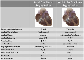
In patients with structurally normal atrioventricular valvular apparatus, functional regurgitation of the mitral or tricuspid valves has been attributed mainly to ventricular dilation and/or dysfunction, through a combination of annulus dilation and tethering of the valve leaflets. The occurrence of functional regurgitation of atrioventricular valves in patients with long-standing persistent atrial fibrillation and atrial dilation but normal ventricular size and function has received much less attention, and its peculiar mechanisms still remain to be understood. This distinct form of functional regurgitation (i.e., "atrial functional regurgitation") may require different treatment and interventional repair approaches than the classical functional regurgitation due to ventricular dilatation and dysfunction ("ventricular functional regurgitation"), and current guideline recommendations do not yet address this distinction. Clarifying the differences in the pathophysiology of atrial functional regurgitation and its management implications is of paramount importance. This review describes briefly the comparative anatomy of mitral and tricuspid apparatus and the pathophysiology and typical echocardiographic features of atrial functional regurgitation compared with ventricular functional regurgitation, as well as the added value of three-dimensional echocardiography as an essential imaging tool to clarify the mechanisms involved in its development.
中文翻译:

房室瓣功能性返流和心房颤动:一个难以捉摸的病理生理学环节,值得进一步关注。
在房室瓣膜结构正常的患者中,二尖瓣或三尖瓣的功能性反流主要归因于瓣膜小叶的瓣膜环扩张和束缚,从而使心室扩张和/或功能障碍。长期持续性房颤和心房扩张但正常的心室大小和功能的患者发生房室瓣膜功能性反流的问题已很少受到关注,其特殊机制仍有待了解。功能性返流的这种独特形式(即“心房功能性返流”)可能由于心室扩张和功能障碍而需要不同于经典功能性返流的治疗和介入修复方法(“ 明确房性功能不全的病理生理学差异及其管理意义至关重要。这篇综述简要描述了二尖瓣和三尖瓣器械的比较解剖结构,以及与室功能性反流相比房性功能性反流的病理生理学和典型的超声心动图特征,以及三维超声心动图作为一种基本成像工具的附加值,以阐明所涉及的机制在发展中。明确房性功能不全的病理生理学差异及其管理意义至关重要。这篇综述简要描述了二尖瓣和三尖瓣器械的比较解剖结构,以及与心室功能性反流相比房功能性反流的病理生理学和典型超声心动图特征,以及三维超声心动图作为阐明相关机制必不可少的成像工具的附加价值在发展中。
更新日期:2019-11-01
中文翻译:
房室瓣功能性返流和心房颤动:一个难以捉摸的病理生理学环节,值得进一步关注。
在房室瓣膜结构正常的患者中,二尖瓣或三尖瓣的功能性反流主要归因于瓣膜小叶的瓣膜环扩张和束缚,从而使心室扩张和/或功能障碍。长期持续性房颤和心房扩张但正常的心室大小和功能的患者发生房室瓣膜功能性反流的问题已很少受到关注,其特殊机制仍有待了解。功能性返流的这种独特形式(即“心房功能性返流”)可能由于心室扩张和功能障碍而需要不同于经典功能性返流的治疗和介入修复方法(“ 明确房性功能不全的病理生理学差异及其管理意义至关重要。这篇综述简要描述了二尖瓣和三尖瓣器械的比较解剖结构,以及与室功能性反流相比房性功能性反流的病理生理学和典型的超声心动图特征,以及三维超声心动图作为一种基本成像工具的附加值,以阐明所涉及的机制在发展中。明确房性功能不全的病理生理学差异及其管理意义至关重要。这篇综述简要描述了二尖瓣和三尖瓣器械的比较解剖结构,以及与心室功能性反流相比房功能性反流的病理生理学和典型超声心动图特征,以及三维超声心动图作为阐明相关机制必不可少的成像工具的附加价值在发展中。


 京公网安备 11010802027423号