当前位置:
X-MOL 学术
›
J. Biophotonics
›
论文详情
Our official English website, www.x-mol.net, welcomes your
feedback! (Note: you will need to create a separate account there.)
Classifying T cell activity in autofluorescence intensity images with convolutional neural networks.
Journal of Biophotonics ( IF 2.0 ) Pub Date : 2019-12-15 , DOI: 10.1002/jbio.201960050 Zijie J Wang 1, 2 , Alex J Walsh 2 , Melissa C Skala 2, 3 , Anthony Gitter 1, 2, 4
Journal of Biophotonics ( IF 2.0 ) Pub Date : 2019-12-15 , DOI: 10.1002/jbio.201960050 Zijie J Wang 1, 2 , Alex J Walsh 2 , Melissa C Skala 2, 3 , Anthony Gitter 1, 2, 4
Affiliation
|
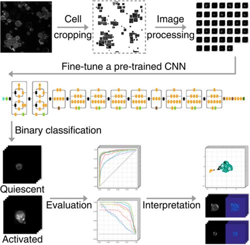
The importance of T cells in immunotherapy has motivated developing technologies to improve therapeutic efficacy. One objective is assessing antigen‐induced T cell activation because only functionally active T cells are capable of killing the desired targets. Autofluorescence imaging can distinguish T cell activity states in a non‐destructive manner by detecting endogenous changes in metabolic co‐enzymes such as NAD(P)H. However, recognizing robust activity patterns is computationally challenging in the absence of exogenous labels. We demonstrate machine learning methods that can accurately classify T cell activity across human donors from NAD(P)H intensity images. Using 8260 cropped single‐cell images from six donors, we evaluate classifiers ranging from traditional models that use previously‐extracted image features to convolutional neural networks (CNNs) pre‐trained on general non‐biological images. Adapting pre‐trained CNNs for the T cell activity classification task provides substantially better performance than traditional models or a simple CNN trained with the autofluorescence images alone. Visualizing the images with dimension reduction provides intuition into why the CNNs achieve higher accuracy than other approaches. Our image processing and classifier training software is available at https://github.com/gitter‐lab/t‐cell‐classification.
中文翻译:

使用卷积神经网络对自发荧光强度图像中的 T 细胞活性进行分类。
T 细胞在免疫治疗中的重要性促使人们开发提高治疗效果的技术。目标之一是评估抗原诱导的 T 细胞激活,因为只有功能活跃的 T 细胞才能杀死所需的靶标。自发荧光成像可以通过检测 NAD(P)H 等代谢辅酶的内源性变化,以非破坏性的方式区分 T 细胞活性状态。然而,在没有外源标签的情况下,识别稳健的活动模式在计算上具有挑战性。我们展示了机器学习方法,可以根据 NAD(P)H 强度图像对人类捐赠者的 T 细胞活性进行准确分类。使用来自 6 个捐赠者的 8260 张裁剪后的单细胞图像,我们评估了从使用先前提取的图像特征的传统模型到在一般非生物图像上预训练的卷积神经网络 (CNN) 的分类器。与传统模型或仅使用自发荧光图像训练的简单 CNN 相比,采用预先训练的 CNN 来执行 T 细胞活动分类任务可提供更好的性能。通过降维对图像进行可视化,可以直观地了解为什么 CNN 比其他方法能实现更高的准确度。我们的图像处理和分类器培训软件可在 https://github.com/gitter-lab/t-cell-classification 上获取。
更新日期:2019-12-15
中文翻译:
使用卷积神经网络对自发荧光强度图像中的 T 细胞活性进行分类。
T 细胞在免疫治疗中的重要性促使人们开发提高治疗效果的技术。目标之一是评估抗原诱导的 T 细胞激活,因为只有功能活跃的 T 细胞才能杀死所需的靶标。自发荧光成像可以通过检测 NAD(P)H 等代谢辅酶的内源性变化,以非破坏性的方式区分 T 细胞活性状态。然而,在没有外源标签的情况下,识别稳健的活动模式在计算上具有挑战性。我们展示了机器学习方法,可以根据 NAD(P)H 强度图像对人类捐赠者的 T 细胞活性进行准确分类。使用来自 6 个捐赠者的 8260 张裁剪后的单细胞图像,我们评估了从使用先前提取的图像特征的传统模型到在一般非生物图像上预训练的卷积神经网络 (CNN) 的分类器。与传统模型或仅使用自发荧光图像训练的简单 CNN 相比,采用预先训练的 CNN 来执行 T 细胞活动分类任务可提供更好的性能。通过降维对图像进行可视化,可以直观地了解为什么 CNN 比其他方法能实现更高的准确度。我们的图像处理和分类器培训软件可在 https://github.com/gitter-lab/t-cell-classification 上获取。


 京公网安备 11010802027423号