Our official English website, www.x-mol.net, welcomes your
feedback! (Note: you will need to create a separate account there.)
Radial somatic F-actin organization affects growth cone dynamics during early neuronal development.
EMBO Reports ( IF 6.5 ) Pub Date : 2019-10-24 , DOI: 10.15252/embr.201947743 Durga Praveen Meka 1 , Robin Scharrenberg 1 , Bing Zhao 1 , Oliver Kobler 2 , Theresa König 1 , Irina Schaefer 1 , Birgit Schwanke 1 , Sergei Klykov 3 , Melanie Richter 1 , Dennis Eggert 4, 5 , Sabine Windhorst 6 , Carlos G Dotti 7 , Michael R Kreutz 8, 9 , Marina Mikhaylova 3 , Froylan Calderon de Anda 1
EMBO Reports ( IF 6.5 ) Pub Date : 2019-10-24 , DOI: 10.15252/embr.201947743 Durga Praveen Meka 1 , Robin Scharrenberg 1 , Bing Zhao 1 , Oliver Kobler 2 , Theresa König 1 , Irina Schaefer 1 , Birgit Schwanke 1 , Sergei Klykov 3 , Melanie Richter 1 , Dennis Eggert 4, 5 , Sabine Windhorst 6 , Carlos G Dotti 7 , Michael R Kreutz 8, 9 , Marina Mikhaylova 3 , Froylan Calderon de Anda 1
Affiliation
|
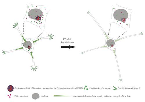
The centrosome is thought to be the major neuronal microtubule-organizing center (MTOC) in early neuronal development, producing microtubules with a radial organization. In addition, albeit in vitro, recent work showed that isolated centrosomes could serve as an actin-organizing center, raising the possibility that neuronal development may, in addition, require a centrosome-based actin radial organization. Here, we report, using super-resolution microscopy and live-cell imaging of cultured rodent neurons, F-actin organization around the centrosome with dynamic F-actin aster-like structures with F-actin fibers extending and retracting actively. Photoactivation/photoconversion experiments and molecular manipulations of F-actin stability reveal a robust flux of somatic F-actin toward the cell periphery. Finally, we show that somatic F-actin intermingles with centrosomal PCM-1 (pericentriolar material 1 protein) satellites. Knockdown of PCM-1 and disruption of centrosomal activity not only affect F-actin dynamics near the centrosome but also in distal growth cones. Collectively, the data show a radial F-actin organization during early neuronal development, which might be a cellular mechanism for providing peripheral regions with a fast and continuous source of actin polymers, hence sustaining initial neuronal development.
中文翻译:

径向体细胞 F-肌动蛋白组织影响早期神经元发育过程中的生长锥动力学。
中心体被认为是早期神经元发育中主要的神经元微管组织中心(MTOC),产生具有放射状组织的微管。此外,尽管是在体外,最近的研究表明,分离的中心体可以作为肌动蛋白组织中心,这增加了神经元发育可能还需要基于中心体的肌动蛋白放射状组织的可能性。在这里,我们使用超分辨率显微镜和培养的啮齿动物神经元的活细胞成像报告,中心体周围的 F-肌动蛋白组织具有动态的 F-肌动蛋白紫苑样结构,其中 F-肌动蛋白纤维主动伸展和缩回。光活化/光转换实验和 F-肌动蛋白稳定性的分子操作揭示了体细胞 F-肌动蛋白向细胞外周的强劲流动。最后,我们证明体细胞 F-肌动蛋白与中心体 PCM-1(中心粒周围物质 1 蛋白)卫星混合。 PCM-1 的敲低和中心体活性的破坏不仅影响中心体附近的 F-肌动蛋白动力学,而且还影响远端生长锥的 F-肌动蛋白动力学。总的来说,数据显示了早期神经元发育过程中的放射状 F-肌动蛋白组织,这可能是为周围区域提供快速且连续的肌动蛋白聚合物来源的细胞机制,从而维持初始神经元发育。
更新日期:2019-12-05
中文翻译:
径向体细胞 F-肌动蛋白组织影响早期神经元发育过程中的生长锥动力学。
中心体被认为是早期神经元发育中主要的神经元微管组织中心(MTOC),产生具有放射状组织的微管。此外,尽管是在体外,最近的研究表明,分离的中心体可以作为肌动蛋白组织中心,这增加了神经元发育可能还需要基于中心体的肌动蛋白放射状组织的可能性。在这里,我们使用超分辨率显微镜和培养的啮齿动物神经元的活细胞成像报告,中心体周围的 F-肌动蛋白组织具有动态的 F-肌动蛋白紫苑样结构,其中 F-肌动蛋白纤维主动伸展和缩回。光活化/光转换实验和 F-肌动蛋白稳定性的分子操作揭示了体细胞 F-肌动蛋白向细胞外周的强劲流动。最后,我们证明体细胞 F-肌动蛋白与中心体 PCM-1(中心粒周围物质 1 蛋白)卫星混合。 PCM-1 的敲低和中心体活性的破坏不仅影响中心体附近的 F-肌动蛋白动力学,而且还影响远端生长锥的 F-肌动蛋白动力学。总的来说,数据显示了早期神经元发育过程中的放射状 F-肌动蛋白组织,这可能是为周围区域提供快速且连续的肌动蛋白聚合物来源的细胞机制,从而维持初始神经元发育。


 京公网安备 11010802027423号