当前位置:
X-MOL 学术
›
STEM CELLS
›
论文详情
Our official English website, www.x-mol.net, welcomes your
feedback! (Note: you will need to create a separate account there.)
SPARC Promotes Self-Renewal of Limbal Epithelial Stem Cells and Ocular Surface Restoration through JNK and p38-MAPK Signaling Pathways
STEM CELLS ( IF 4.0 ) Pub Date : 2019-11-19 , DOI: 10.1002/stem.3100 Jing Zhu 1 , Le-Yi Wang 1 , Chong-Yun Li 1 , Jia-Yin Wu 1 , Yu-Ting Zhang 1 , Kun-Peng Pang 1 , Yan Wei 2 , Li-Qun Du 1 , Mei Liu 1 , Xin-Yi Wu 1
STEM CELLS ( IF 4.0 ) Pub Date : 2019-11-19 , DOI: 10.1002/stem.3100 Jing Zhu 1 , Le-Yi Wang 1 , Chong-Yun Li 1 , Jia-Yin Wu 1 , Yu-Ting Zhang 1 , Kun-Peng Pang 1 , Yan Wei 2 , Li-Qun Du 1 , Mei Liu 1 , Xin-Yi Wu 1
Affiliation
|
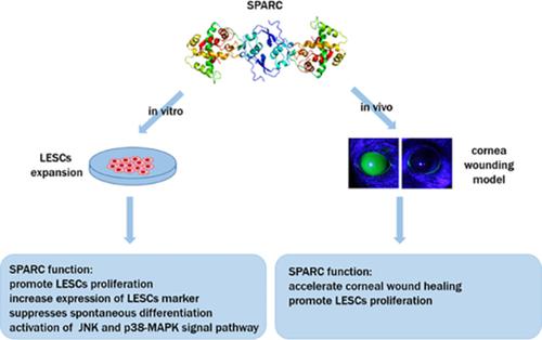
The purpose of this study was to investigate the effects of secreted protein acidic and rich in cysteine (SPARC) on the maintenance of limbal epithelial stem cell (LESC) stemness and restoration of ocular surface. To determine the suitable concentration of SPARC for LESC culture, the marker expression, mitogenic effect, and holoclone‐forming capacity of LESCs treated with different concentrations of SPARC were analyzed. To investigate the mechanism of SPARC's action on the preservation of LESCs stemness, the phosphorylation of related signaling pathways was evaluated by Western blotting. A corneal wound model was established to verify the function of SPARC in ocular surface repair. Consecutive subculturing, colony‐forming efficiency, immunofluorescence, and 5‐ethynyl‐2‐deoxyuridine incorporation assays indicated that 1 μg/mL SPARC was a suitable concentration to stimulate LESC proliferation and preserve their proliferative potential. Compared with a control group, 1 μg/mL SPARC effectively increased the expression of ABCG‐2, Bmi‐1, and Ki67, while decreasing that of CK3/12. The mitogenic effect of SPARC on LESCs was found to be mediated by the phosphorylation of c‐Jun N‐terminal kinase (JNK) and p38‐MAPK signaling pathways, whereas the inhibitors of JNK and p38 MAPK reduced the marker expression and mitogenic capacity of LESCs. In a corneal injury model, SPARC facilitated corneal epithelial wound healing and promoted the proliferation of p63α‐positive cells both in the limbus and in the epithelial healing front. SPARC promotes proliferation while suppressing spontaneous differentiation of LESCs through JNK and p38‐MAPK signaling pathways, suggesting that SPARC is a promising factor for the improvement of LESCs culture in vitro and in vivo.
中文翻译:

SPARC 通过 JNK 和 p38-MAPK 信号通路促进边缘上皮干细胞的自我更新和眼表修复
本研究的目的是研究分泌蛋白酸性和富含半胱氨酸(SPARC)对角膜缘上皮干细胞(LESC)干细胞的维持和眼表修复的影响。为了确定适合 LESC 培养的 SPARC 浓度,分析了用不同浓度 SPARC 处理的 LESC 的标记表达、促有丝分裂作用和全克隆形成能力。为了研究 SPARC 对 LESCs 干性保护的作用机制,通过蛋白质印迹评估相关信号通路的磷酸化。建立角膜创面模型以验证SPARC在眼表修复中的作用。连续传代、集落形成效率、免疫荧光、和 5-乙炔基-2-脱氧尿苷掺入试验表明,1 μg/mL SPARC 是刺激 LESC 增殖并保持其增殖潜力的合适浓度。与对照组相比,1 μg/mL SPARC 有效增加 ABCG-2、Bmi-1 和 Ki67 的表达,同时降低 CK3/12 的表达。SPARC 对 LESCs 的促有丝分裂作用被发现是由 c-Jun N-末端激酶 (JNK) 和 p38-MAPK 信号通路的磷酸化介导的,而 JNK 和 p38 MAPK 的抑制剂降低了 LESCs 的标志物表达和有丝分裂能力. 在角膜损伤模型中,SPARC 促进角膜上皮伤口愈合并促进角膜缘和上皮愈合前沿 p63α 阳性细胞的增殖。
更新日期:2019-11-19
中文翻译:
SPARC 通过 JNK 和 p38-MAPK 信号通路促进边缘上皮干细胞的自我更新和眼表修复
本研究的目的是研究分泌蛋白酸性和富含半胱氨酸(SPARC)对角膜缘上皮干细胞(LESC)干细胞的维持和眼表修复的影响。为了确定适合 LESC 培养的 SPARC 浓度,分析了用不同浓度 SPARC 处理的 LESC 的标记表达、促有丝分裂作用和全克隆形成能力。为了研究 SPARC 对 LESCs 干性保护的作用机制,通过蛋白质印迹评估相关信号通路的磷酸化。建立角膜创面模型以验证SPARC在眼表修复中的作用。连续传代、集落形成效率、免疫荧光、和 5-乙炔基-2-脱氧尿苷掺入试验表明,1 μg/mL SPARC 是刺激 LESC 增殖并保持其增殖潜力的合适浓度。与对照组相比,1 μg/mL SPARC 有效增加 ABCG-2、Bmi-1 和 Ki67 的表达,同时降低 CK3/12 的表达。SPARC 对 LESCs 的促有丝分裂作用被发现是由 c-Jun N-末端激酶 (JNK) 和 p38-MAPK 信号通路的磷酸化介导的,而 JNK 和 p38 MAPK 的抑制剂降低了 LESCs 的标志物表达和有丝分裂能力. 在角膜损伤模型中,SPARC 促进角膜上皮伤口愈合并促进角膜缘和上皮愈合前沿 p63α 阳性细胞的增殖。


 京公网安备 11010802027423号