当前位置:
X-MOL 学术
›
BBA Bioenerg.
›
论文详情
Our official English website, www.x-mol.net, welcomes your
feedback! (Note: you will need to create a separate account there.)
Pigment-protein complexes are organized into stable microdomains in cyanobacterial thylakoids.
Biochimica et Biophysica Acta (BBA) - Bioenergetics ( IF 3.4 ) Pub Date : 2019-07-22 , DOI: 10.1016/j.bbabio.2019.07.008 A Strašková 1 , G Steinbach 1 , G Konert 1 , E Kotabová 1 , J Komenda 1 , M Tichý 1 , R Kaňa 1
Biochimica et Biophysica Acta (BBA) - Bioenergetics ( IF 3.4 ) Pub Date : 2019-07-22 , DOI: 10.1016/j.bbabio.2019.07.008 A Strašková 1 , G Steinbach 1 , G Konert 1 , E Kotabová 1 , J Komenda 1 , M Tichý 1 , R Kaňa 1
Affiliation
|
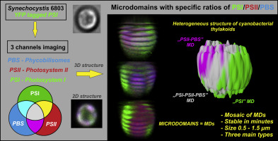
Thylakoids are the place of the light-photosynthetic reactions. To gain maximal efficiency, these reactions are conditional to proper pigment-pigment and protein-protein interactions. In higher plants thylakoids, the interactions lead to a lateral asymmetry in localization of protein complexes (i.e. granal/stromal thylakoids) that have been defined as a domain-like structures characteristic by different biochemical composition and function (Albertsson P-Å. 2001,Trends Plant Science 6: 349-354). We explored this complex organization of thylakoid pigment-proteins at single cell level in the cyanobacterium Synechocystis sp. PCC 6803. Our 3D confocal images captured heterogeneous distribution of all main photosynthetic pigment-protein complexes (PPCs), Photosystem I (fluorescently tagged by YFP), Photosystem II and Phycobilisomes. The acquired images depicted cyanobacterial thylakoid membrane as a stable, mosaic-like structure formed by microdomains (MDs). These microcompartments are of sub-micrometer in sizes (~0.5-1.5 μm), typical by particular PPCs ratios and importantly without full segregation of observed complexes. The most prevailing MD is represented by MD with high Photosystem I content which allows also partial separation of Photosystems like in higher plants thylakoids. We assume that MDs stability (in minutes) provides optimal conditions for efficient excitation/electron transfer. The cyanobacterial MDs thus define thylakoid membrane organization as a system controlled by co-localization of three main PPCs leading to formation of thylakoid membrane mosaic. This organization might represent evolutional and functional precursor for the granal/stromal spatial heterogeneity in photosystems that is typical for higher plant thylakoids.
中文翻译:

色素-蛋白质复合物在蓝细菌类囊体中被组织成稳定的微区。
类固醇是光-光合作用反应的地方。为了获得最大效率,这些反应必须以适当的颜料-色素和蛋白质-蛋白质相互作用为条件。在高等植物类囊体中,相互作用导致蛋白质复合物(即粒状/基质类囊体)的定位出现横向不对称性,该复合物已被定义为具有不同生化组成和功能的结构域样结构(AlbertssonP-Å。2001,趋势植物科学6:349-354)。我们探讨了蓝藻Synechocystis sp。中单细胞水平类囊体色素蛋白的这种复杂的组织。PCC6803。我们的3D共聚焦图像捕获了所有主要光合色素蛋白复合物(PPC),光系统I(由YFP荧光标记),光系统II和藻胆体的异质分布。所获取的图像将蓝细菌类囊体膜描述为由微区(MDs)形成的稳定,马赛克状结构。这些微隔室的尺寸为亚微米级(〜0.5-1.5μm),通常通过特定的PPC比率实现,而且重要的是,观察到的复合物没有完全隔离。最普遍的MD以具有较高Photosystem I含量的MD表示,这也允许像高等植物类囊体中的Photosystems进行部分分离。我们假设MD的稳定性(以分钟为单位)为有效的激发/电子转移提供了最佳条件。因此,蓝细菌MD将类囊体膜组织定义为由三个主要PPC的共定位控制的系统,从而导致类囊体膜镶嵌的形成。
更新日期:2019-07-22
中文翻译:
色素-蛋白质复合物在蓝细菌类囊体中被组织成稳定的微区。
类固醇是光-光合作用反应的地方。为了获得最大效率,这些反应必须以适当的颜料-色素和蛋白质-蛋白质相互作用为条件。在高等植物类囊体中,相互作用导致蛋白质复合物(即粒状/基质类囊体)的定位出现横向不对称性,该复合物已被定义为具有不同生化组成和功能的结构域样结构(AlbertssonP-Å。2001,趋势植物科学6:349-354)。我们探讨了蓝藻Synechocystis sp。中单细胞水平类囊体色素蛋白的这种复杂的组织。PCC6803。我们的3D共聚焦图像捕获了所有主要光合色素蛋白复合物(PPC),光系统I(由YFP荧光标记),光系统II和藻胆体的异质分布。所获取的图像将蓝细菌类囊体膜描述为由微区(MDs)形成的稳定,马赛克状结构。这些微隔室的尺寸为亚微米级(〜0.5-1.5μm),通常通过特定的PPC比率实现,而且重要的是,观察到的复合物没有完全隔离。最普遍的MD以具有较高Photosystem I含量的MD表示,这也允许像高等植物类囊体中的Photosystems进行部分分离。我们假设MD的稳定性(以分钟为单位)为有效的激发/电子转移提供了最佳条件。因此,蓝细菌MD将类囊体膜组织定义为由三个主要PPC的共定位控制的系统,从而导致类囊体膜镶嵌的形成。


 京公网安备 11010802027423号