当前位置:
X-MOL 学术
›
J. Food Drug Anal.
›
论文详情
Our official English website, www.x-mol.net, welcomes your feedback! (Note: you will need to create a separate account there.)
Involvement of prohibitin 1 and prohibitin 2 upregulation in cBSA-induced podocyte cytotoxicity
Journal of Food and Drug Analysis ( IF 3.6 ) Pub Date : 2020-01-01 , DOI: 10.1016/j.jfda.2019.09.003 Heng-Hsiung Wu , Chao-Jung Chen , Pei-Yu Lin , Yu-Huei Liu
Journal of Food and Drug Analysis ( IF 3.6 ) Pub Date : 2020-01-01 , DOI: 10.1016/j.jfda.2019.09.003 Heng-Hsiung Wu , Chao-Jung Chen , Pei-Yu Lin , Yu-Huei Liu
|
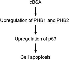
Membranous nephropathy (MN) is the most common cause of nephrotic syndrome in adults, when not effectively treated. The aim of this study was to discover new targets for the diagnosis and treatment of MN. A reliable mouse model of MN was used by the administration of cationic bovine serum albumin (cBSA). Mice with MN exhibited proteinuria, histopathological changes, and accumulation of immune complexes in the glomerular basement membrane. Label-free proteomics analysis was performed to identify changes in protein expression, and the overexpressed proteins were evaluated. There were 273 proteins that showed significantly different expression in mice with MN, as compared to the controls. String analysis showed that functions related to cellular catabolic processes were downregulated in MN. Among the differentially expressed proteins, prohibitin 1 (PHB1) and prohibitin 2 (PHB2) were upregulated in the kidneys of mice with MN, as demonstrated by immunohistochemistry (IHC), and this upregulation was observed in both the tubular cells and glomeruli. Both shRNA-mediated knockdown of PHB1 or PHB2 inhibited tumor suppressor p53 expression and significantly promoted podocyte proliferation. In addition, both PHB1 and PHB2 were responsible for cBSA-induced cytotoxicity. Microarray analysis further revealed that the upregulation of PHB1 and PHB2 may be due to a blockage of proteasome activity. These data demonstrate that the upregulation of PHB2 is involved in cBSA-mediated podocyte cytotoxicity, which may lead to MN development.
中文翻译:

抑制素 1 和抑制素 2 上调参与 cBSA 诱导的足细胞细胞毒性
如果未得到有效治疗,膜性肾病 (MN) 是成人肾病综合征的最常见原因。本研究的目的是发现诊断和治疗 MN 的新靶点。通过施用阳离子牛血清白蛋白 (cBSA) 使用了可靠的 MN 小鼠模型。患有 MN 的小鼠表现出蛋白尿、组织病理学变化和免疫复合物在肾小球基底膜中的积累。进行无标记蛋白质组学分析以鉴定蛋白质表达的变化,并评估过表达的蛋白质。与对照组相比,有 273 种蛋白质在 MN 小鼠中表现出显着不同的表达。字符串分析表明,与细胞分解代谢过程相关的功能在 MN 中被下调。在差异表达的蛋白质中,正如免疫组织化学 (IHC) 所证明的那样,抑制素 1 (PHB1) 和抑制素 2 (PHB2) 在 MN 小鼠的肾脏中上调,并且在肾小管细胞和肾小球中均观察到这种上调。shRNA 介导的 PHB1 或 PHB2 敲低均抑制肿瘤抑制因子 p53 的表达并显着促进足细胞增殖。此外,PHB1 和 PHB2 都负责 cBSA 诱导的细胞毒性。微阵列分析进一步表明,PHB1 和 PHB2 的上调可能是由于蛋白酶体活性受阻所致。这些数据表明,PHB2 的上调涉及 cBSA 介导的足细胞细胞毒性,这可能导致 MN 发展。并且在肾小管细胞和肾小球中都观察到了这种上调。shRNA 介导的 PHB1 或 PHB2 敲低均抑制肿瘤抑制因子 p53 的表达并显着促进足细胞增殖。此外,PHB1 和 PHB2 都负责 cBSA 诱导的细胞毒性。微阵列分析进一步表明,PHB1 和 PHB2 的上调可能是由于蛋白酶体活性受阻所致。这些数据表明,PHB2 的上调涉及 cBSA 介导的足细胞细胞毒性,这可能导致 MN 发展。并且在肾小管细胞和肾小球中都观察到了这种上调。shRNA 介导的 PHB1 或 PHB2 敲低均抑制肿瘤抑制因子 p53 的表达并显着促进足细胞增殖。此外,PHB1 和 PHB2 都负责 cBSA 诱导的细胞毒性。微阵列分析进一步表明,PHB1 和 PHB2 的上调可能是由于蛋白酶体活性受阻所致。这些数据表明,PHB2 的上调涉及 cBSA 介导的足细胞细胞毒性,这可能导致 MN 发展。微阵列分析进一步表明,PHB1 和 PHB2 的上调可能是由于蛋白酶体活性受阻所致。这些数据表明,PHB2 的上调涉及 cBSA 介导的足细胞细胞毒性,这可能导致 MN 发展。微阵列分析进一步表明,PHB1 和 PHB2 的上调可能是由于蛋白酶体活性受阻所致。这些数据表明,PHB2 的上调涉及 cBSA 介导的足细胞细胞毒性,这可能导致 MN 发展。
更新日期:2020-01-01
中文翻译:
抑制素 1 和抑制素 2 上调参与 cBSA 诱导的足细胞细胞毒性
如果未得到有效治疗,膜性肾病 (MN) 是成人肾病综合征的最常见原因。本研究的目的是发现诊断和治疗 MN 的新靶点。通过施用阳离子牛血清白蛋白 (cBSA) 使用了可靠的 MN 小鼠模型。患有 MN 的小鼠表现出蛋白尿、组织病理学变化和免疫复合物在肾小球基底膜中的积累。进行无标记蛋白质组学分析以鉴定蛋白质表达的变化,并评估过表达的蛋白质。与对照组相比,有 273 种蛋白质在 MN 小鼠中表现出显着不同的表达。字符串分析表明,与细胞分解代谢过程相关的功能在 MN 中被下调。在差异表达的蛋白质中,正如免疫组织化学 (IHC) 所证明的那样,抑制素 1 (PHB1) 和抑制素 2 (PHB2) 在 MN 小鼠的肾脏中上调,并且在肾小管细胞和肾小球中均观察到这种上调。shRNA 介导的 PHB1 或 PHB2 敲低均抑制肿瘤抑制因子 p53 的表达并显着促进足细胞增殖。此外,PHB1 和 PHB2 都负责 cBSA 诱导的细胞毒性。微阵列分析进一步表明,PHB1 和 PHB2 的上调可能是由于蛋白酶体活性受阻所致。这些数据表明,PHB2 的上调涉及 cBSA 介导的足细胞细胞毒性,这可能导致 MN 发展。并且在肾小管细胞和肾小球中都观察到了这种上调。shRNA 介导的 PHB1 或 PHB2 敲低均抑制肿瘤抑制因子 p53 的表达并显着促进足细胞增殖。此外,PHB1 和 PHB2 都负责 cBSA 诱导的细胞毒性。微阵列分析进一步表明,PHB1 和 PHB2 的上调可能是由于蛋白酶体活性受阻所致。这些数据表明,PHB2 的上调涉及 cBSA 介导的足细胞细胞毒性,这可能导致 MN 发展。并且在肾小管细胞和肾小球中都观察到了这种上调。shRNA 介导的 PHB1 或 PHB2 敲低均抑制肿瘤抑制因子 p53 的表达并显着促进足细胞增殖。此外,PHB1 和 PHB2 都负责 cBSA 诱导的细胞毒性。微阵列分析进一步表明,PHB1 和 PHB2 的上调可能是由于蛋白酶体活性受阻所致。这些数据表明,PHB2 的上调涉及 cBSA 介导的足细胞细胞毒性,这可能导致 MN 发展。微阵列分析进一步表明,PHB1 和 PHB2 的上调可能是由于蛋白酶体活性受阻所致。这些数据表明,PHB2 的上调涉及 cBSA 介导的足细胞细胞毒性,这可能导致 MN 发展。微阵列分析进一步表明,PHB1 和 PHB2 的上调可能是由于蛋白酶体活性受阻所致。这些数据表明,PHB2 的上调涉及 cBSA 介导的足细胞细胞毒性,这可能导致 MN 发展。


 京公网安备 11010802027423号