Our official English website, www.x-mol.net, welcomes your
feedback! (Note: you will need to create a separate account there.)
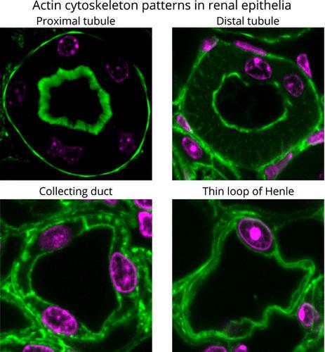
Identification and classification of epithelial cells in nephron segments by actin cytoskeleton patterns.
The FEBS Journal ( IF 5.5 ) Pub Date : 2019-10-12 , DOI: 10.1111/febs.15088 Girishkumar Kaitholil Kumaran 1 , Israel Hanukoglu 1
The FEBS Journal ( IF 5.5 ) Pub Date : 2019-10-12 , DOI: 10.1111/febs.15088 Girishkumar Kaitholil Kumaran 1 , Israel Hanukoglu 1
Affiliation
|
The basic functional unit in a kidney is the nephron, which is a long and morphologically segmented tubule. The nephron begins with a cluster of capillaries called glomerulus through which the blood is filtered into the Bowman's space. The filtrate flows through the nephron segments. During this flow, electrolytes and solutes are reabsorbed by channels and transport systems into the capillaries wrapped around the nephron. Many questions related to renal function focus on identifying the sites of expression of these systems. In this study, we mapped whole kidney sections by confocal microscopic imaging of fluorescent phalloidin, which binds to actin filaments. In tile scans (composed of hundreds of images) of these sections, the cortex and the medullary regions (outer and inner stripes of the outer medulla, and inner medulla) could be easily identified by their cytoskeletal patterns. At a higher resolution, we identified distinct features of the actin cytoskeleton in the apical, basal, and lateral borders of the cells. These features could be used to identify segments of a nephron (the proximal tubule, thin and thick segments of Henle's loop, and distal tubule), the collecting duct system, the papillary ducts in the papilla, and the urothelium that covers the pelvis. To verify our findings, we used additional markers, including aquaporin isoforms, cytokeratin 8-18, and WGA lectin. This study highlights the power of high-resolution confocal microscopy for identifying specific cell types using the simple probe of F-actin-binding phalloidin.
中文翻译:

通过肌动蛋白细胞骨架模式鉴定和分类肾单位中的上皮细胞。
肾脏的基本功能单位是肾单位,它是一个长且在形态上分段的肾小管。肾单位开始于一团称为肾小球的毛细血管,血液通过毛细血管过滤进入鲍曼氏空间。滤液流过肾单位段。在此流动过程中,电解质和溶质被通道和传输系统重新吸收到包裹在肾单位周围的毛细管中。与肾脏功能有关的许多问题都集中在确定这些系统的表达部位上。在这项研究中,我们通过共聚焦显微镜成像荧光鬼笔环肽(与肌动蛋白丝结合)绘制了整个肾脏切片。在这些部分的平铺扫描(由数百张图像组成)中,皮质和髓质区域(外部髓质的外部和内部条纹)和内侧髓质)可以很容易地通过它们的细胞骨架模式来识别。在更高的分辨率,我们确定了肌动蛋白细胞骨架在细胞的顶端,基底和外侧边界的不同特征。这些功能可用于识别肾单位的各个部分(近端小管,亨利氏环的薄而厚的部分以及远端小管),收集管系统,乳头中的乳头管以及覆盖骨盆的尿道上皮。为了验证我们的发现,我们使用了其他标记,包括水通道蛋白同工型,细胞角蛋白8-18和WGA凝集素。这项研究突出了高分辨率共聚焦显微镜使用F-肌动蛋白结合鬼笔环肽的简单探针鉴定特定细胞类型的能力。我们确定了肌动蛋白细胞骨架在细胞的顶,基和外侧边界中的独特特征。这些功能可用于识别肾单位的各个部分(近端小管,亨利氏环的薄而厚的部分以及远端小管),收集管系统,乳头中的乳头管以及覆盖骨盆的尿道上皮。为了验证我们的发现,我们使用了其他标记,包括水通道蛋白同工型,细胞角蛋白8-18和WGA凝集素。这项研究突出了高分辨率共聚焦显微镜使用F-肌动蛋白结合鬼笔环肽的简单探针鉴定特定细胞类型的能力。我们确定了肌动蛋白细胞骨架在细胞的顶,基和外侧边界中的独特特征。这些功能可用于识别肾单位的各个部分(近端小管,亨利氏环的薄而厚的部分以及远端小管),收集管系统,乳头中的乳头管以及覆盖骨盆的尿道上皮。为了验证我们的发现,我们使用了其他标记,包括水通道蛋白同工型,细胞角蛋白8-18和WGA凝集素。这项研究突出了高分辨率共聚焦显微镜使用F-肌动蛋白结合鬼笔环肽的简单探针鉴定特定细胞类型的能力。和远端小管),收集导管系统,乳头中的乳头导管以及覆盖骨盆的尿道上皮。为了验证我们的发现,我们使用了其他标记,包括水通道蛋白同工型,细胞角蛋白8-18和WGA凝集素。这项研究突出了高分辨率共聚焦显微镜使用F-肌动蛋白结合鬼笔环肽的简单探针鉴定特定细胞类型的能力。和远端小管),收集导管系统,乳头中的乳头导管以及覆盖骨盆的尿道上皮。为了验证我们的发现,我们使用了其他标记,包括水通道蛋白同工型,细胞角蛋白8-18和WGA凝集素。这项研究突出了高分辨率共聚焦显微镜使用F-肌动蛋白结合鬼笔环肽的简单探针鉴定特定细胞类型的能力。
更新日期:2020-03-16
中文翻译:
通过肌动蛋白细胞骨架模式鉴定和分类肾单位中的上皮细胞。
肾脏的基本功能单位是肾单位,它是一个长且在形态上分段的肾小管。肾单位开始于一团称为肾小球的毛细血管,血液通过毛细血管过滤进入鲍曼氏空间。滤液流过肾单位段。在此流动过程中,电解质和溶质被通道和传输系统重新吸收到包裹在肾单位周围的毛细管中。与肾脏功能有关的许多问题都集中在确定这些系统的表达部位上。在这项研究中,我们通过共聚焦显微镜成像荧光鬼笔环肽(与肌动蛋白丝结合)绘制了整个肾脏切片。在这些部分的平铺扫描(由数百张图像组成)中,皮质和髓质区域(外部髓质的外部和内部条纹)和内侧髓质)可以很容易地通过它们的细胞骨架模式来识别。在更高的分辨率,我们确定了肌动蛋白细胞骨架在细胞的顶端,基底和外侧边界的不同特征。这些功能可用于识别肾单位的各个部分(近端小管,亨利氏环的薄而厚的部分以及远端小管),收集管系统,乳头中的乳头管以及覆盖骨盆的尿道上皮。为了验证我们的发现,我们使用了其他标记,包括水通道蛋白同工型,细胞角蛋白8-18和WGA凝集素。这项研究突出了高分辨率共聚焦显微镜使用F-肌动蛋白结合鬼笔环肽的简单探针鉴定特定细胞类型的能力。我们确定了肌动蛋白细胞骨架在细胞的顶,基和外侧边界中的独特特征。这些功能可用于识别肾单位的各个部分(近端小管,亨利氏环的薄而厚的部分以及远端小管),收集管系统,乳头中的乳头管以及覆盖骨盆的尿道上皮。为了验证我们的发现,我们使用了其他标记,包括水通道蛋白同工型,细胞角蛋白8-18和WGA凝集素。这项研究突出了高分辨率共聚焦显微镜使用F-肌动蛋白结合鬼笔环肽的简单探针鉴定特定细胞类型的能力。我们确定了肌动蛋白细胞骨架在细胞的顶,基和外侧边界中的独特特征。这些功能可用于识别肾单位的各个部分(近端小管,亨利氏环的薄而厚的部分以及远端小管),收集管系统,乳头中的乳头管以及覆盖骨盆的尿道上皮。为了验证我们的发现,我们使用了其他标记,包括水通道蛋白同工型,细胞角蛋白8-18和WGA凝集素。这项研究突出了高分辨率共聚焦显微镜使用F-肌动蛋白结合鬼笔环肽的简单探针鉴定特定细胞类型的能力。和远端小管),收集导管系统,乳头中的乳头导管以及覆盖骨盆的尿道上皮。为了验证我们的发现,我们使用了其他标记,包括水通道蛋白同工型,细胞角蛋白8-18和WGA凝集素。这项研究突出了高分辨率共聚焦显微镜使用F-肌动蛋白结合鬼笔环肽的简单探针鉴定特定细胞类型的能力。和远端小管),收集导管系统,乳头中的乳头导管以及覆盖骨盆的尿道上皮。为了验证我们的发现,我们使用了其他标记,包括水通道蛋白同工型,细胞角蛋白8-18和WGA凝集素。这项研究突出了高分辨率共聚焦显微镜使用F-肌动蛋白结合鬼笔环肽的简单探针鉴定特定细胞类型的能力。


 京公网安备 11010802027423号