当前位置:
X-MOL 学术
›
J. Biophotonics
›
论文详情
Our official English website, www.x-mol.net, welcomes your
feedback! (Note: you will need to create a separate account there.)
Localization of PD-L1 on single cancer cells by iSERS microscopy with Au/Au core/satellite nanoparticles.
Journal of Biophotonics ( IF 2.0 ) Pub Date : 2020-01-01 , DOI: 10.1002/jbio.201960034 Elzbieta Stepula 1 , Matthias König 1 , Xin-Ping Wang 1 , Janina Levermann 2 , Tobias Schimming 3 , Sabine Kasimir-Bauer 2 , Bastian Schilling 3, 4 , Sebastian Schlücker 1
Journal of Biophotonics ( IF 2.0 ) Pub Date : 2020-01-01 , DOI: 10.1002/jbio.201960034 Elzbieta Stepula 1 , Matthias König 1 , Xin-Ping Wang 1 , Janina Levermann 2 , Tobias Schimming 3 , Sabine Kasimir-Bauer 2 , Bastian Schilling 3, 4 , Sebastian Schlücker 1
Affiliation
|
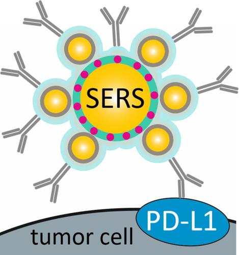
Programmed cell death‐ligand 1 (PD‐L1) is an important predictive biomarker. The detection of PD‐L1 can be crucial for patients with advanced cancer where the use of immunotherapy is considered. Here, we demonstrate the use of immuno‐SERS microscopy (iSERS) for localizing PD‐L1 on single cancer SkBr‐3 cells. A central advantage of iSERS is that the disturbing autofluorescence from cells and tissues can be efficiently minimized by red to near‐infrared laser excitation. In this study we employed Au/Au core/satellite nanoparticles as SERS nanotags because of their remarkable signal brightness and colloidal stability upon red laser excitation. False‐color iSERS images of the positive and negative controls clearly reveal the specific localization of PD‐L1 with SERS nanotag‐labeled antibodies.
中文翻译:

使用 Au/Au 核心/卫星纳米颗粒通过 iSERS 显微镜定位单个癌细胞上的 PD-L1。
程序性细胞死亡配体 1 (PD-L1) 是一种重要的预测生物标志物。PD-L1 的检测对于考虑使用免疫疗法的晚期癌症患者至关重要。在这里,我们演示了使用免疫 SERS 显微镜 (iSERS) 将 PD-L1 定位在单个癌症 SkBr-3 细胞上。iSERS 的一个核心优势是,可以通过红色至近红外激光激发有效地最大程度地减少来自细胞和组织的干扰性自发荧光。在这项研究中,我们采用 Au/Au 核/卫星纳米粒子作为 SERS 纳米标签,因为它们在红色激光激发下具有显着的信号亮度和胶体稳定性。阳性和阴性对照的假彩色 iSERS 图像清楚地揭示了带有 SERS 纳米标签标记抗体的 PD-L1 的特异性定位。
更新日期:2020-01-01
中文翻译:
使用 Au/Au 核心/卫星纳米颗粒通过 iSERS 显微镜定位单个癌细胞上的 PD-L1。
程序性细胞死亡配体 1 (PD-L1) 是一种重要的预测生物标志物。PD-L1 的检测对于考虑使用免疫疗法的晚期癌症患者至关重要。在这里,我们演示了使用免疫 SERS 显微镜 (iSERS) 将 PD-L1 定位在单个癌症 SkBr-3 细胞上。iSERS 的一个核心优势是,可以通过红色至近红外激光激发有效地最大程度地减少来自细胞和组织的干扰性自发荧光。在这项研究中,我们采用 Au/Au 核/卫星纳米粒子作为 SERS 纳米标签,因为它们在红色激光激发下具有显着的信号亮度和胶体稳定性。阳性和阴性对照的假彩色 iSERS 图像清楚地揭示了带有 SERS 纳米标签标记抗体的 PD-L1 的特异性定位。


 京公网安备 11010802027423号