当前位置:
X-MOL 学术
›
Toxicol. Res.
›
论文详情
Our official English website, www.x-mol.net, welcomes your
feedback! (Note: you will need to create a separate account there.)
Biogenic Aspergillus tubingensis silver nanoparticles' in vitro effects on human umbilical vein endothelial cells, normal human fibroblasts, HEPG2, and Galleria mellonella.
Toxicology Research ( IF 2.2 ) Pub Date : 2019-10-10 , DOI: 10.1039/c9tx00091g Cristiane Angélica Ottoni 1 , Durvanei Augusto Maria 2 , Priscila Jane Romano de Oliveira Gonçalves 3 , Welington Luiz de Araújo 3 , Ana Olívia de Souza 2
Toxicology Research ( IF 2.2 ) Pub Date : 2019-10-10 , DOI: 10.1039/c9tx00091g Cristiane Angélica Ottoni 1 , Durvanei Augusto Maria 2 , Priscila Jane Romano de Oliveira Gonçalves 3 , Welington Luiz de Araújo 3 , Ana Olívia de Souza 2
Affiliation
|
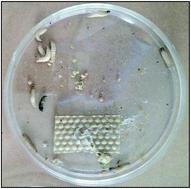
Silver nanoparticles (AgNPs) are widely incorporated into different hygiene, personal care, and healthcare products. However, few studies have been undertaken to determine the effects of biogenic AgNPs on human health. The effect of biosynthesized AgNPs using the fungus Aspergillus tubingensis culture was evaluated on human umbilical vein endothelial cells (HUVECs), normal human fibroblasts (FN1), human hepatoma cells (HEPG2) and a Galleria mellonella model. HUVECs were more susceptible to biogenic AgNPs than normal fibroblasts FN1 and intense cytotoxicity was observed only for very high concentrations at and above 2.5 μM for both cells. Normal human fibroblasts FN1 exposed to AgNPs for 24 h showed viability of 98.83 ± 8.40% and 94.86 ± 5.50% for 1.25 and 2.5 μM, respectively. At 5 and 10 μM, related to the control, an increase in cell viability was observed being 112.66 ± 9.94% and 117.86 ± 8.86%, respectively. Similar results were obtained for treatment for 48 and 72 h. At 1.25, 2.5, 5 and 10 μM of AgNPs, at 24 h, HUVECs showed 51.34 ± 7.47%, 27.01 ± 5.77%, 26.00 ± 3.03% and 27.64 ± 5.85% of viability, respectively. No alteration in cell distribution among different cycle phases was observed after HUVEC and normal fibroblast FN1 exposure to AgNPs from 0.01 to 1 μM for 24, 48 and 72 h. Based on the clonogenic assay, nanoparticles successfully inhibited HEPG2 cell proliferation when exposed to concentrations up to 1 μM. In addition to that, AgNPs did not induce senescence and no morphological alteration was observed by scanning electron microscopy on the endothelial cells. In the larvae of the wax moth, Galleria mellonella, a model for toxicity, AgNPs showed no significant effects, which corroborates to the safety of their use in mammalian cells. These results demonstrate that the use of A. tubingensis AgNPs is a promising biotechnological approach and these AgNPs can be applied in several biomedical situations.
中文翻译:

生物源塔宾曲霉银纳米颗粒对人脐静脉内皮细胞、正常人成纤维细胞、HEPG2 和大蜡螟的体外影响。
银纳米颗粒 (AgNP) 广泛应用于不同的卫生、个人护理和保健产品中。然而,很少有研究来确定生物纳米银颗粒对人类健康的影响。使用真菌塔宾曲霉培养物生物合成的 AgNP 对人脐静脉内皮细胞 (HUVEC)、正常人成纤维细胞 (FN1)、人肝癌细胞 (HEPG2) 和大蜡螟模型的效果进行了评估。 HUVEC 比正常成纤维细胞 FN1 更容易受到生物 AgNP 的影响,并且两种细胞仅在 2.5 μM 及以上的极高浓度下才观察到强烈的细胞毒性。正常人成纤维细胞 FN1 暴露于 AgNP 24 小时,对于 1.25 和 2.5 μM 的活性分别为 98.83 ± 8.40% 和 94.86 ± 5.50%。在 5 和 10 μM 浓度下,与对照相比,观察到细胞活力分别增加了 112.66 ± 9.94% 和 117.86 ± 8.86%。处理 48 小时和 72 小时也获得了类似的结果。在 1.25、2.5、5 和 10 μM AgNP 下,24 小时时,HUVEC 的活力分别为 51.34 ± 7.47%、27.01 ± 5.77%、26.00 ± 3.03% 和 27.64 ± 5.85%。 HUVEC 和正常成纤维细胞 FN1 暴露于 0.01 至 1 μM AgNP 24、48 和 72 小时后,未观察到不同周期阶段的细胞分布变化。根据克隆形成实验,纳米粒子在浓度高达 1 μM 时成功抑制 HEPG2 细胞增殖。除此之外,AgNPs 不会诱导衰老,并且通过扫描电子显微镜观察内皮细胞没有观察到形态变化。在蜡蛾幼虫(一种毒性模型)中,AgNPs 没有表现出明显的作用,这证实了它们在哺乳动物细胞中使用的安全性。 这些结果表明,使用塔宾金丝猴 AgNP 是一种有前途的生物技术方法,这些 AgNP 可应用于多种生物医学情况。
更新日期:2019-10-10
中文翻译:
生物源塔宾曲霉银纳米颗粒对人脐静脉内皮细胞、正常人成纤维细胞、HEPG2 和大蜡螟的体外影响。
银纳米颗粒 (AgNP) 广泛应用于不同的卫生、个人护理和保健产品中。然而,很少有研究来确定生物纳米银颗粒对人类健康的影响。使用真菌塔宾曲霉培养物生物合成的 AgNP 对人脐静脉内皮细胞 (HUVEC)、正常人成纤维细胞 (FN1)、人肝癌细胞 (HEPG2) 和大蜡螟模型的效果进行了评估。 HUVEC 比正常成纤维细胞 FN1 更容易受到生物 AgNP 的影响,并且两种细胞仅在 2.5 μM 及以上的极高浓度下才观察到强烈的细胞毒性。正常人成纤维细胞 FN1 暴露于 AgNP 24 小时,对于 1.25 和 2.5 μM 的活性分别为 98.83 ± 8.40% 和 94.86 ± 5.50%。在 5 和 10 μM 浓度下,与对照相比,观察到细胞活力分别增加了 112.66 ± 9.94% 和 117.86 ± 8.86%。处理 48 小时和 72 小时也获得了类似的结果。在 1.25、2.5、5 和 10 μM AgNP 下,24 小时时,HUVEC 的活力分别为 51.34 ± 7.47%、27.01 ± 5.77%、26.00 ± 3.03% 和 27.64 ± 5.85%。 HUVEC 和正常成纤维细胞 FN1 暴露于 0.01 至 1 μM AgNP 24、48 和 72 小时后,未观察到不同周期阶段的细胞分布变化。根据克隆形成实验,纳米粒子在浓度高达 1 μM 时成功抑制 HEPG2 细胞增殖。除此之外,AgNPs 不会诱导衰老,并且通过扫描电子显微镜观察内皮细胞没有观察到形态变化。在蜡蛾幼虫(一种毒性模型)中,AgNPs 没有表现出明显的作用,这证实了它们在哺乳动物细胞中使用的安全性。 这些结果表明,使用塔宾金丝猴 AgNP 是一种有前途的生物技术方法,这些 AgNP 可应用于多种生物医学情况。


 京公网安备 11010802027423号