Cell Death & Disease ( IF 9 ) Pub Date : 2019-09-26 , DOI: 10.1038/s41419-019-1913-6 Marc Oria 1 , Soner Duru 1 , Rebeca L Figueira 1, 2 , Federico Scorletti 1, 3 , Lucas E Turner 4, 5 , Irati Fernandez-Alonso 1 , Alejandra Fernandez-Martin 1, 6 , Mario Marotta 1, 6 , Lourenco Sbragia 1, 2 , Aimen F Shaaban 4, 5 , Jose L Peiro 1
|
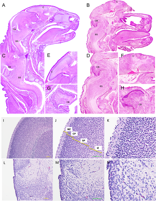
Exencephaly/anencephaly is one of the leading causes of neonatal mortality and the most extreme open neural tube defect with no current treatments and limited mechanistic understanding. We hypothesized that exencephaly leads to a local neurodegenerative process in the brain exposed to the amniotic fluid as well as diffuse degeneration in other encephalic areas and the spinal cord. To evaluate the consequences of in utero neural tissue exposure, brain and spinal cord samples from E17 exencephalic murine fetuses (maternal intraperitoneal administration of valproic acid at E8) were analyzed and compared to controls and saline-injected shams (n = 11/group). Expression of apoptosis and senescence genes (p53, p21, p16, Rbl2, Casp3, Casp9) was determined by qRT-PCR and protein expression analyzed by western blot. Apoptosis was measured by TUNEL assay and PI/AV flow cytometry. Valproic acid at E8 induced exencephaly in 22% of fetuses. At E17 the fetuses exhibited the characteristic absence of cranial bones. The brain structures from exencephalic fetuses demonstrated a loss of layers in cortical regions and a complete loss of structural organization in the olfactory bulb, hippocampus, dental gyrus and septal cortex. E17 fetuses had reduced expression of NeuN, GFAP and Oligodendrocytes in the brain with primed microglia. Intrinsic apoptotic activation (p53, Caspase9 and 3) was upregulated and active Caspase3 localized to the layer of brain exposed to the amniotic fluid. Senescence via p21-Rbl2 was increased in the brain and in the spinal cord at the lamina I-II of the somatosensory dorsal horn. The current study characterizes CNS alterations in murine exencephaly and demonstrates that degeneration due to intrinsic apoptosis and senescence occurs in the directly exposed brain but also remotely in the spinal cord.
中文翻译:
细胞坏死,内在的细胞凋亡和衰老有助于先天性颅骨分裂症小鼠模型中的自发性向无脑性发展。
脑无力/无脑是新生儿死亡和最极端的开放性神经管缺损的主要原因之一,目前尚无治疗方法,并且对机械的了解有限。我们假设,自由运动导致暴露于羊水的大脑局部神经变性过程,以及其他脑区和脊髓的弥漫性变性。为了评估子宫内神经组织暴露的后果,分析了E17脑外鼠胎儿(在E8时腹膜内给予丙戊酸的孕妇腹腔注射)的脑和脊髓样本,并将其与对照组和注射盐水的sha子(n = 11 /组)。通过qRT-PCR确定凋亡和衰老基因(p53,p21,p16,Rbl2,Casp3,Casp9)的表达,并通过蛋白质印迹分析蛋白质表达。通过TUNEL测定和PI / AV流式细胞术测量细胞凋亡。E8处的丙戊酸在22%的胎儿中诱导出自发性。在E17时,胎儿表现出没有颅骨的特征。脑外胎儿的大脑结构显示皮质区域的层丢失,嗅球,海马,齿状回和中隔皮质的结构组织完全丧失。E17胎儿的初生小胶质细胞在大脑中的NeuN,GFAP和少突胶质细胞表达减少。内在凋亡激活(p53,Caspase9和3)被上调,而活跃的Caspase3定位于暴露于羊水的大脑层。在体感背角I-II层的大脑和脊髓中,通过p21-Rbl2引起的衰老增加了。目前的研究表征了小鼠中枢神经系统中CNS的改变,并证明了由于固有的凋亡和衰老引起的变性发生在直接暴露的大脑中,也发生在远程的脊髓中。


 京公网安备 11010802027423号