Our official English website, www.x-mol.net, welcomes your
feedback! (Note: you will need to create a separate account there.)
Extracellular matrix in ascending aortic aneurysms and dissections - What we learn from decellularization and scanning electron microscopy.
PLOS ONE ( IF 2.9 ) Pub Date : 2019-03-18 , DOI: 10.1371/journal.pone.0213794 Teresa Mimler 1 , Clemens Nebert 1 , Eva Eichmair 1 , Birgitta Winter 1 , Thomas Aschacher 2 , Marie-Elisabeth Stelzmueller 2 , Martin Andreas 2 , Marek Ehrlich 2 , Guenther Laufer 2 , Barbara Messner 1
PLOS ONE ( IF 2.9 ) Pub Date : 2019-03-18 , DOI: 10.1371/journal.pone.0213794 Teresa Mimler 1 , Clemens Nebert 1 , Eva Eichmair 1 , Birgitta Winter 1 , Thomas Aschacher 2 , Marie-Elisabeth Stelzmueller 2 , Martin Andreas 2 , Marek Ehrlich 2 , Guenther Laufer 2 , Barbara Messner 1
Affiliation
|
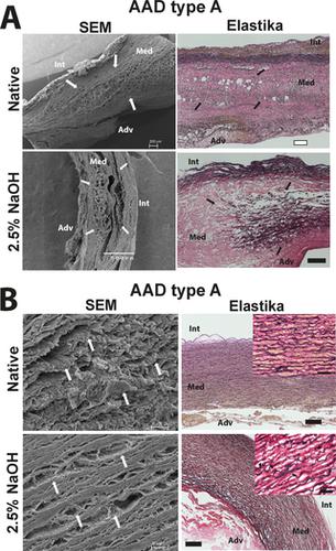
Pathological impairment of elastic fiber and other extracellular matrix (ECM) components are described for the aortic media of ascending thoracic aortic aneurysms (aTAA) but the exact pathological impairment of the structure and its degree still needs further investigations. To evaluate the quantity and quality of elastic fiber sheets and other ECM structures (e.g. collagen), cells were removed from different types of aneurysmal tissues (tricuspid aortic valve [TAV] associated-, bicuspid aortic valve [BAV] associated-aneurysmal tissue and acute aortic dissections [AAD]) using 2.5% sodium hydroxide (NaOH) and compared to decellularized control aortic tissue. Likewise, native tissue has been analysed. To evaluate the 2D- (histological evaluation, fluorescence- and auto-fluorescence based staining methods) and the 3D structure (scanning electron microscopic [SEM] examination) of the medial layer we first analysed for a successful decellularization. After proving for successful decellularization, we quantified the amount of elastic fiber sheets, elastin and other ECM components including collagen. Aside from clearly visible focal elastic fiber loss in TAV-aTAA tissue, decellularization resulted in reduction of elastic fiber auto-fluorescence properties, which is perhaps an indication from a disease-related qualitative impairment of elastic fibers, visible only after contact with the alkaline solution. Likewise, the loss of collagen amount in BAV-aTAA and TAV-aTAA tissue (compared to non-decellularized tissue) after contact with NaOH indicates a prior disease-associated impairment of collagen. Although the amount of ECM was not changed in type A dissection tissue, detailed electron microscopic evaluation revealed changes in ECM quality, which worsened after contact with alkaline solution but were not visible after histological analyses. Apart from the improved observation of the samples using electron microscopy, contact of aneurysmal and dissected tissue with the alkaline decellularization solution revealed potential disease related changes in ECM quality which can partly be connected to already published data, but have to be proven by further studies.
中文翻译:

升主动脉瘤和夹层中的细胞外基质-我们从脱细胞和扫描电子显微镜学到的东西。
弹性纤维和其他细胞外基质(ECM)组件的病理损伤已被描述为升主动脉瘤(aTAA)的主动脉介质,但确切的病理损伤及其结构程度仍需进一步研究。为了评估弹性纤维片和其他ECM结构(例如胶原蛋白)的数量和质量,从不同类型的动脉瘤组织(三尖瓣主动脉[TAV]相关,双尖瓣主动脉[BAV]相关的非动脉瘤组织和急性主动脉夹层[AAD])使用2.5%氢氧化钠(NaOH)进行比较,并与脱细胞的对照主动脉组织进行比较。同样,已经分析了天然组织。要评估2D-(组织学评估,我们首先分析了内侧层的3D结构(基于扫描荧光和自发荧光的染色方法)和3D结构(扫描电子显微镜[SEM]检查),以成功进行脱细胞。在证明成功脱细胞后,我们量化了弹性纤维片,弹性蛋白和其他ECM成分(包括胶原蛋白)的数量。除了在TAV-aTAA组织中清晰可见的局灶性弹性纤维损失外,脱细胞作用还导致弹性纤维自发荧光性能降低,这可能是疾病相关的弹性纤维定性损伤的征兆,仅在与碱性溶液接触后可见。同样地,与NaOH接触后,BAV-aTAA和TAV-aTAA组织(与未脱细胞的组织相比)中胶原蛋白量的损失表明胶原与疾病相关。尽管在A型解剖组织中ECM的量没有变化,但是详细的电子显微镜评估显示ECM的质量发生了变化,与碱性溶液接触后会变差,但在组织学分析后看不到。除了使用电子显微镜更好地观察样品外,动脉瘤和解剖组织与碱性脱细胞溶液的接触还揭示了潜在的疾病相关的ECM质量变化,这可以部分与已经发表的数据联系起来,但必须通过进一步的研究加以证明。详细的电子显微镜评估显示ECM质量的变化,与碱性溶液接触后变差,但在组织学分析后不可见。除了使用电子显微镜更好地观察样品外,动脉瘤和解剖组织与碱性脱细胞溶液的接触还揭示了潜在的疾病相关的ECM质量变化,这可以部分与已经发表的数据联系起来,但必须通过进一步的研究加以证明。详细的电子显微镜评估显示ECM质量的变化,与碱性溶液接触后变差,但在组织学分析后不可见。除了使用电子显微镜更好地观察样品外,动脉瘤和解剖组织与碱性脱细胞溶液的接触还揭示了潜在的疾病相关的ECM质量变化,这可以部分与已经发表的数据联系起来,但必须通过进一步的研究加以证明。
更新日期:2019-03-19
中文翻译:
升主动脉瘤和夹层中的细胞外基质-我们从脱细胞和扫描电子显微镜学到的东西。
弹性纤维和其他细胞外基质(ECM)组件的病理损伤已被描述为升主动脉瘤(aTAA)的主动脉介质,但确切的病理损伤及其结构程度仍需进一步研究。为了评估弹性纤维片和其他ECM结构(例如胶原蛋白)的数量和质量,从不同类型的动脉瘤组织(三尖瓣主动脉[TAV]相关,双尖瓣主动脉[BAV]相关的非动脉瘤组织和急性主动脉夹层[AAD])使用2.5%氢氧化钠(NaOH)进行比较,并与脱细胞的对照主动脉组织进行比较。同样,已经分析了天然组织。要评估2D-(组织学评估,我们首先分析了内侧层的3D结构(基于扫描荧光和自发荧光的染色方法)和3D结构(扫描电子显微镜[SEM]检查),以成功进行脱细胞。在证明成功脱细胞后,我们量化了弹性纤维片,弹性蛋白和其他ECM成分(包括胶原蛋白)的数量。除了在TAV-aTAA组织中清晰可见的局灶性弹性纤维损失外,脱细胞作用还导致弹性纤维自发荧光性能降低,这可能是疾病相关的弹性纤维定性损伤的征兆,仅在与碱性溶液接触后可见。同样地,与NaOH接触后,BAV-aTAA和TAV-aTAA组织(与未脱细胞的组织相比)中胶原蛋白量的损失表明胶原与疾病相关。尽管在A型解剖组织中ECM的量没有变化,但是详细的电子显微镜评估显示ECM的质量发生了变化,与碱性溶液接触后会变差,但在组织学分析后看不到。除了使用电子显微镜更好地观察样品外,动脉瘤和解剖组织与碱性脱细胞溶液的接触还揭示了潜在的疾病相关的ECM质量变化,这可以部分与已经发表的数据联系起来,但必须通过进一步的研究加以证明。详细的电子显微镜评估显示ECM质量的变化,与碱性溶液接触后变差,但在组织学分析后不可见。除了使用电子显微镜更好地观察样品外,动脉瘤和解剖组织与碱性脱细胞溶液的接触还揭示了潜在的疾病相关的ECM质量变化,这可以部分与已经发表的数据联系起来,但必须通过进一步的研究加以证明。详细的电子显微镜评估显示ECM质量的变化,与碱性溶液接触后变差,但在组织学分析后不可见。除了使用电子显微镜更好地观察样品外,动脉瘤和解剖组织与碱性脱细胞溶液的接触还揭示了潜在的疾病相关的ECM质量变化,这可以部分与已经发表的数据联系起来,但必须通过进一步的研究加以证明。


 京公网安备 11010802027423号