Our official English website, www.x-mol.net, welcomes your
feedback! (Note: you will need to create a separate account there.)
The intracellular Ca2+ concentration is elevated in cardiomyocytes differentiated from hiPSCs derived from a Duchenne muscular dystrophy patient.
PLOS ONE ( IF 2.9 ) Pub Date : 2019-03-15 , DOI: 10.1371/journal.pone.0213768 Fumitoshi Tsurumi 1 , Shiro Baba 1 , Daisuke Yoshinaga 1 , Katsutsugu Umeda 1 , Takuya Hirata 1 , Junko Takita 1 , Toshio Heike 1
PLOS ONE ( IF 2.9 ) Pub Date : 2019-03-15 , DOI: 10.1371/journal.pone.0213768 Fumitoshi Tsurumi 1 , Shiro Baba 1 , Daisuke Yoshinaga 1 , Katsutsugu Umeda 1 , Takuya Hirata 1 , Junko Takita 1 , Toshio Heike 1
Affiliation
|
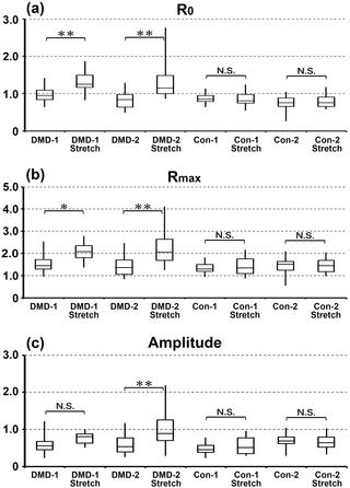
Duchenne muscular dystrophy (DMD) is the most common and severe form of muscular dystrophy. The major symptoms of this condition are walking difficulties, dyspnea caused by progressive skeletal muscle weakness, and cardiomyopathy. Recent advances in ventilator support devices have dramatically decreased mortality caused by respiratory distress. Consequently, cardiomyopathy resulting in heart failure is currently the major cause of death among DMD patients. One mechanism by which skeletal muscle is damaged in DMD patients involves elevation of the intracellular Ca2+ concentration. By contrast, the mechanisms underlying the development of cardiomyopathy are unclear. To investigate this, we examined the intracellular Ca2+ concentration and calcium transients in cardiomyocytes differentiated from human induced pluripotent stem cells (hiPSCs). hiPSCs were derived from a DMD patient (DMD-hiPSCs), in whom exon 44 of the gene encoding dystrophin was deleted, and from his parents (control-hiPSCs), who did not carry this mutation. The intracellular Ca2+ concentration was measured using the fluorescent indicator indo-1. The fluorescence ratio (410/490 nm) of indo-1 at rest (R0), the peak of this ratio (Rmax), and the amplitude (Rmax-R0) were significantly higher in cardiomyocytes differentiated from DMD-hiPSCs than in those differentiated from control-hiPSCs. Moreover, mechanical stretching significantly increased the intracellular Ca2+ concentration in cardiomyocytes differentiated from DMD-hiPSCs, but not in those differentiated from control-hiPSCs. These findings indicate that elevation of the intracellular Ca2+ concentration can cause cardiac damage leading to cardiomyopathy in DMD patients.
中文翻译:

心肌细胞中的细胞内Ca2 +浓度升高,该细胞与源自杜兴氏肌营养不良症患者的hiPSC分化。
杜氏肌营养不良症(DMD)是最常见和最严重的肌肉营养不良形式。这种情况的主要症状是行走困难,进行性骨骼肌无力引起的呼吸困难和心肌病。呼吸机支撑装置的最新进展已大大降低了因呼吸窘迫而导致的死亡率。因此,导致心力衰竭的心肌病目前是DMD患者中主要的死亡原因。DMD患者骨骼肌受损的一种机制涉及细胞内Ca2 +浓度的升高。相比之下,心肌病发展的潜在机制尚不清楚。为了对此进行研究,我们检查了与人诱导多能干细胞(hiPSCs)分化的心肌细胞的细胞内Ca2 +浓度和钙瞬变。hiPSC来源于DMD患者(DMD-hiPSC),后者的编码肌营养不良蛋白的基因的第44外显子被删除,其父母则来自未携带此突变的双亲(control-hiPSC)。使用荧光指示剂indo-1测量细胞内Ca2 +浓度。从DMD-hiPSC分化而来的心肌细胞中,静止时indo-1的荧光比率(R0)的荧光比率(410/490 nm),该比率的峰值(Rmax)和振幅(Rmax-R0)显着高于分化的心肌细胞。来自control-hiPSC。此外,机械拉伸显着增加了从DMD-hiPSCs分化而来的心肌细胞中的细胞内Ca2 +浓度,但在与对照-hiPSCs分化而来的心肌细胞中却没有。
更新日期:2019-03-17
中文翻译:
心肌细胞中的细胞内Ca2 +浓度升高,该细胞与源自杜兴氏肌营养不良症患者的hiPSC分化。
杜氏肌营养不良症(DMD)是最常见和最严重的肌肉营养不良形式。这种情况的主要症状是行走困难,进行性骨骼肌无力引起的呼吸困难和心肌病。呼吸机支撑装置的最新进展已大大降低了因呼吸窘迫而导致的死亡率。因此,导致心力衰竭的心肌病目前是DMD患者中主要的死亡原因。DMD患者骨骼肌受损的一种机制涉及细胞内Ca2 +浓度的升高。相比之下,心肌病发展的潜在机制尚不清楚。为了对此进行研究,我们检查了与人诱导多能干细胞(hiPSCs)分化的心肌细胞的细胞内Ca2 +浓度和钙瞬变。hiPSC来源于DMD患者(DMD-hiPSC),后者的编码肌营养不良蛋白的基因的第44外显子被删除,其父母则来自未携带此突变的双亲(control-hiPSC)。使用荧光指示剂indo-1测量细胞内Ca2 +浓度。从DMD-hiPSC分化而来的心肌细胞中,静止时indo-1的荧光比率(R0)的荧光比率(410/490 nm),该比率的峰值(Rmax)和振幅(Rmax-R0)显着高于分化的心肌细胞。来自control-hiPSC。此外,机械拉伸显着增加了从DMD-hiPSCs分化而来的心肌细胞中的细胞内Ca2 +浓度,但在与对照-hiPSCs分化而来的心肌细胞中却没有。


 京公网安备 11010802027423号