npj Regenerative Medicine ( IF 6.4 ) Pub Date : 2018-02-09 , DOI: 10.1038/s41536-018-0042-7 M B Albro 1, 2, 3 , M S Bergholt 1, 2, 3 , J P St-Pierre 1, 2, 3 , A Vinals Guitart 1, 2, 3 , H M Zlotnick 1, 2, 3 , E G Evita 1, 2, 3 , M M Stevens 1, 2, 3
|
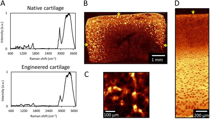
Articular cartilage possesses a remarkable, mechanically-robust extracellular matrix (ECM) that is organized and distributed throughout the tissue to resist physiologic strains and provide low friction during articulation. The ability to characterize the make-up and distribution of the cartilage ECM is critical to both understand the process by which articular cartilage undergoes disease-related degeneration and to develop novel tissue repair strategies to restore tissue functionality. However, the ability to quantitatively measure the spatial distribution of cartilage ECM constituents throughout the tissue has remained a major challenge. In this experimental investigation, we assessed the analytical ability of Raman micro-spectroscopic imaging to semi-quantitatively measure the distribution of the major ECM constituents in cartilage tissues. Raman spectroscopic images were acquired of two distinct cartilage tissue types that possess large spatial ECM gradients throughout their depth: native articular cartilage explants and large engineered cartilage tissue constructs. Spectral acquisitions were processed via multivariate curve resolution to decompose the “fingerprint” range spectra (800–1800 cm−1) to the component spectra of GAG, collagen, and water, giving rise to the depth dependent concentration profile of each constituent throughout the tissues. These Raman spectroscopic acquired-profiles exhibited strong agreement with profiles independently acquired via direct biochemical assaying of spatial tissue sections. Further, we harness this spectroscopic technique to evaluate local heterogeneities through the depth of cartilage. This work represents a powerful analytical validation of the accuracy of Raman spectroscopic imaging measurements of the spatial distribution of biochemical components in a biological tissue and shows that it can be used as a valuable tool for quantitatively measuring the distribution and organization of ECM constituents in native and engineered cartilage tissue specimens.
中文翻译:
拉曼光谱成像用于量化天然软骨和工程软骨中的深度依赖性和局部异质性
关节软骨拥有卓越的、机械坚固的细胞外基质 (ECM),它组织并分布在整个组织中,以抵抗生理应变并在关节活动期间提供低摩擦。表征软骨 ECM 的组成和分布的能力对于了解关节软骨经历疾病相关退化的过程以及开发新的组织修复策略以恢复组织功能至关重要。然而,定量测量整个组织中软骨 ECM 成分的空间分布的能力仍然是一个重大挑战。在这项实验研究中,我们评估了拉曼显微光谱成像的分析能力,以半定量测量软骨组织中主要 ECM 成分的分布。获得了两种不同的软骨组织类型的拉曼光谱图像,它们在整个深度上具有大的空间 ECM 梯度:天然关节软骨外植体和大型工程软骨组织构建体。通过多元曲线分辨率处理光谱采集,将“指纹”范围光谱 (800–1800 cm -1 ) 分解为 GAG、胶原蛋白和水的成分光谱,从而产生整个组织中每种成分的深度依赖浓度分布。这些拉曼光谱获得的轮廓与通过空间组织切片的直接生化分析独立获得的轮廓表现出强烈的一致性。此外,我们利用这种光谱技术通过软骨深度评估局部异质性。 这项工作代表了对生物组织中生化成分空间分布的拉曼光谱成像测量准确性的有力分析验证,并表明它可以用作定量测量天然和组织中 ECM 成分的分布和组织的有价值的工具。工程软骨组织标本。


 京公网安备 11010802027423号