当前位置:
X-MOL 学术
›
Contrast Media Mol. Imaging
›
论文详情
Our official English website, www.x-mol.net, welcomes your
feedback! (Note: you will need to create a separate account there.)
Dual nano‐sized contrast agents in PET/MRI: a systematic review
Contrast Media & Molecular Imaging Pub Date : 2017-01-19 , DOI: 10.1002/cmmi.1719 Afsaneh Lahooti 1 , Saeed Sarkar 1 , Sophie Laurent 2, 3 , Saeed Shanehsazzadeh 2
Contrast Media & Molecular Imaging Pub Date : 2017-01-19 , DOI: 10.1002/cmmi.1719 Afsaneh Lahooti 1 , Saeed Sarkar 1 , Sophie Laurent 2, 3 , Saeed Shanehsazzadeh 2
Affiliation
|
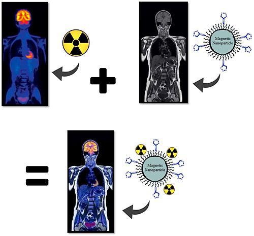
Nowadays molecular imaging plays a vital role in achieving a successful targeted and personalized treatment. Hence, the approach of combining two or more medical imaging modalities was developed. The objective of this review is to systematically compare recent dual contrast agents in Positron Emission Tomography (PET)/Magnetic Resonance Imaging (MRI) and in some cases Single photon emission computed tomography (SPECT)/MRI in terms of some their characteristics, such as tumor uptake, and reticuloendothelial system uptake (especially liver) and their relaxivity rates for early detection of primary cancer tumor. To the best of our knowledge, this is the first systematic and integrated overview of this field. Two reviewers individually directed the systematic review search using PubMed, MEDLINE and Google Scholar. Two other reviewers directed quality assessment, using the criteria checklist from the CAMARADES (Collaborative Approach to Meta‐Analysis and Review of Animal Data from Experimental Studies) tool, and differences were resolved by consensus. After reviewing all 49 studies, we concluded that a size range of 20–200 nm can be used for molecular imaging, although it is better to try to achieve as small a size as it is possible. Also, small nanoparticles with a hydrophilic coating and positive charge are suitable as a T2 contrast agent. According to our selected data, the most successful dual probes in terms of high targeting were with an average size of 40 nm, PEGylated using peptides as a biomarker and radiolabeled with copper 64 and gallium 68. Copyright © 2017 John Wiley & Sons, Ltd.
中文翻译:

PET / MRI中的双重纳米级造影剂:系统综述
如今,分子成像在实现成功的靶向和个性化治疗中起着至关重要的作用。因此,开发了组合两个或更多个医学成像模态的方法。这篇综述的目的是系统地比较正电子发射断层扫描(PET)/磁共振成像(MRI)和某些情况下单光子发射计算机断层扫描(SPECT)/ MRI的最新双重造影剂的某些特性,例如肿瘤摄取和网状内皮系统摄取(尤其是肝脏)及其松弛率,可早期发现原发癌肿瘤。据我们所知,这是该领域的第一个系统的综合概述。两名评论者分别使用PubMed,MEDLINE和Google Scholar指导了系统的评论搜索。其他两名审稿人使用CAMARADES(实验研究的动物数据的荟萃分析和审阅协作方法)工具中的标准清单指导质量评估,并通过共识解决了差异。回顾了所有49项研究后,我们得出结论,分子成像可以使用20-200 nm的尺寸范围,尽管最好尝试尽可能减小尺寸。同样,具有亲水涂层和正电荷的小纳米颗粒也适合作为T 尽管最好尝试尽可能减小尺寸。同样,具有亲水涂层和正电荷的小纳米颗粒也适合作为T 尽管最好尝试尽可能减小尺寸。同样,具有亲水涂层和正电荷的小纳米颗粒也适合作为T2造影剂。根据我们选择的数据,就高靶向而言,最成功的双探针平均大小为40 nm,使用肽作为生物标记物进行聚乙二醇化,并用铜64和镓68进行放射性标记。版权所有©2017 John Wiley&Sons,Ltd.
更新日期:2017-01-19
中文翻译:
PET / MRI中的双重纳米级造影剂:系统综述
如今,分子成像在实现成功的靶向和个性化治疗中起着至关重要的作用。因此,开发了组合两个或更多个医学成像模态的方法。这篇综述的目的是系统地比较正电子发射断层扫描(PET)/磁共振成像(MRI)和某些情况下单光子发射计算机断层扫描(SPECT)/ MRI的最新双重造影剂的某些特性,例如肿瘤摄取和网状内皮系统摄取(尤其是肝脏)及其松弛率,可早期发现原发癌肿瘤。据我们所知,这是该领域的第一个系统的综合概述。两名评论者分别使用PubMed,MEDLINE和Google Scholar指导了系统的评论搜索。其他两名审稿人使用CAMARADES(实验研究的动物数据的荟萃分析和审阅协作方法)工具中的标准清单指导质量评估,并通过共识解决了差异。回顾了所有49项研究后,我们得出结论,分子成像可以使用20-200 nm的尺寸范围,尽管最好尝试尽可能减小尺寸。同样,具有亲水涂层和正电荷的小纳米颗粒也适合作为T 尽管最好尝试尽可能减小尺寸。同样,具有亲水涂层和正电荷的小纳米颗粒也适合作为T 尽管最好尝试尽可能减小尺寸。同样,具有亲水涂层和正电荷的小纳米颗粒也适合作为T2造影剂。根据我们选择的数据,就高靶向而言,最成功的双探针平均大小为40 nm,使用肽作为生物标记物进行聚乙二醇化,并用铜64和镓68进行放射性标记。版权所有©2017 John Wiley&Sons,Ltd.


 京公网安备 11010802027423号