当前位置:
X-MOL 学术
›
Contrast Media Mol. Imaging
›
论文详情
Our official English website, www.x-mol.net, welcomes your
feedback! (Note: you will need to create a separate account there.)
A novel Tc‐99 m and fluorescence labeled peptide as a multimodal imaging agent for targeting angiogenesis in a murine tumor model
Contrast Media & Molecular Imaging Pub Date : 2016-10-14 , DOI: 10.1002/cmmi.1714 Myoung Hyoun Kim 1 , Chang Guhn Kim 1 , Seul-Gi Kim 2 , Dae-Weung Kim 1, 2
Contrast Media & Molecular Imaging Pub Date : 2016-10-14 , DOI: 10.1002/cmmi.1714 Myoung Hyoun Kim 1 , Chang Guhn Kim 1 , Seul-Gi Kim 2 , Dae-Weung Kim 1, 2
Affiliation
|
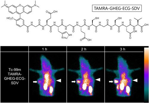
The serine–aspartic acid–valine (SDV) peptide binds specifically to integrin αVβ3. In the present study, we successfully developed a TAMRA–GHEG–ECG–SDV peptide labeled with both Tc‐99 m and TAMRA to target the integrin αVβ3 of tumor cells; furthermore, we evaluated the diagnostic performance of Tc‐99 m TAMRA–GHEG–ECG–SDV as a dual‐modality imaging agent for tumor of the murine model. TAMRA–GHEG–ECG–SDV was synthesized using Fmoc solid‐phase peptide synthesis. Radiolabeling of TAMRA–GHEG–ECG–SDV with Tc‐99 m was done using ligand exchange methods. Labeling stability and cytotoxicity studies were performed. Gamma camera imaging, biodistribution and ex vivo imaging studies were performed in murine models with HT‐1080 and HT‐29 tumors. A tumor tissue slide was prepared and analyzed using confocal microscopy. After radiolabeling procedures with Tc‐99 m, the Tc‐99 m TAMRA–GHEG–ECG–SDV complexes were prepared in high yield (>99%). In the gamma camera imaging study, a substantial uptake of Tc‐99 m TAMRA–GHEG–ECG–SDV into HT‐1080 tumor (integrin αVβ3 positive) and low uptake of Tc‐99 m TAMRA–GHEG–ECG–SDV into HT‐29 tumor (integrin αVβ3 negative) were demonstrated. A competition study revealed that HT‐1080 tumor uptake was effectively blocked by the co‐injection of an excess concentration of SDV. Specific uptake of Tc‐99 m TAMRA–GHEG–ECG–SDV was confirmed by biodistribution, ex vivo imaging and confocal microscopy studies. Our in vivo and in vitro studies revealed substantial uptake of Tc‐99 m TAMRA–GHEG–ECG–SDV in the integrin αVβ3‐positive tumor. Tc‐99 m TAMRA–GHEG–ECG–SDV could be a good candidate for a dual‐modality imaging agent targeting tumor angiogenesis. Copyright © 2016 John Wiley & Sons, Ltd.
中文翻译:

一种新型Tc-99 m和荧光标记的肽作为多模态成像剂,可靶向鼠肿瘤模型中的血管生成
丝氨酸-天冬氨酸-缬氨酸(SDV)肽特异性结合整合素α V β 3。在本研究中,我们成功地开发标记有两个锝-99 m和TAMRA一个TAMRA-GHEG-ECG-SDV肽靶向整合素α V β 3的肿瘤细胞; 此外,我们评估了Tc-99 m TAMRA–GHEG–ECG–SDV作为鼠模型肿瘤的双峰显像剂的诊断性能。TAMRA–GHEG–ECG–SDV是使用Fmoc固相肽合成法合成的。使用配体交换方法对TAM-99m进行TAMRA–GHEG–ECG–SDV的放射性标记。进行了标记稳定性和细胞毒性研究。伽马相机成像,生物分布和离体在具有HT-1080和HT-29肿瘤的小鼠模型中进行了影像学研究。制备肿瘤组织玻片并使用共聚焦显微镜分析。用Tc-99 m进行放射性标记后,可以高产率(> 99%)制备Tc-99 m TAMRA-GHEG-ECG-SDV复合物。在γ相机成像研究,TC-99米TAMRA-GHEG-ECG-SDV的实质摄取到HT-1080肿瘤(整合素α V β 3阳性)和TC-99米TAMRA-GHEG-ECG-SDV的低摄取到HT-29肿瘤(整合素α V β 3阴性)进行了论证。一项竞争研究表明,共注射过量浓度的SDV可有效阻止HT-1080肿瘤的摄取。通过生物分布证实了Tc-99 m TAMRA–GHEG–ECG–SDV的特异性摄取,离体成像和共聚焦显微镜研究。我们的体内和体外研究表明在整合素α锝99米TAMRA-GHEG-ECG-SDV的大量摄取V β 3阳性肿瘤。Tc-99 m TAMRA–GHEG–ECG–SDV可能是靶向肿瘤血管生成的双峰成像剂的良好候选者。版权所有©2016 John Wiley&Sons,Ltd.
更新日期:2016-10-14
中文翻译:
一种新型Tc-99 m和荧光标记的肽作为多模态成像剂,可靶向鼠肿瘤模型中的血管生成
丝氨酸-天冬氨酸-缬氨酸(SDV)肽特异性结合整合素α V β 3。在本研究中,我们成功地开发标记有两个锝-99 m和TAMRA一个TAMRA-GHEG-ECG-SDV肽靶向整合素α V β 3的肿瘤细胞; 此外,我们评估了Tc-99 m TAMRA–GHEG–ECG–SDV作为鼠模型肿瘤的双峰显像剂的诊断性能。TAMRA–GHEG–ECG–SDV是使用Fmoc固相肽合成法合成的。使用配体交换方法对TAM-99m进行TAMRA–GHEG–ECG–SDV的放射性标记。进行了标记稳定性和细胞毒性研究。伽马相机成像,生物分布和离体在具有HT-1080和HT-29肿瘤的小鼠模型中进行了影像学研究。制备肿瘤组织玻片并使用共聚焦显微镜分析。用Tc-99 m进行放射性标记后,可以高产率(> 99%)制备Tc-99 m TAMRA-GHEG-ECG-SDV复合物。在γ相机成像研究,TC-99米TAMRA-GHEG-ECG-SDV的实质摄取到HT-1080肿瘤(整合素α V β 3阳性)和TC-99米TAMRA-GHEG-ECG-SDV的低摄取到HT-29肿瘤(整合素α V β 3阴性)进行了论证。一项竞争研究表明,共注射过量浓度的SDV可有效阻止HT-1080肿瘤的摄取。通过生物分布证实了Tc-99 m TAMRA–GHEG–ECG–SDV的特异性摄取,离体成像和共聚焦显微镜研究。我们的体内和体外研究表明在整合素α锝99米TAMRA-GHEG-ECG-SDV的大量摄取V β 3阳性肿瘤。Tc-99 m TAMRA–GHEG–ECG–SDV可能是靶向肿瘤血管生成的双峰成像剂的良好候选者。版权所有©2016 John Wiley&Sons,Ltd.


 京公网安备 11010802027423号