当前位置:
X-MOL 学术
›
Contrast Media Mol. Imaging
›
论文详情
Our official English website, www.x-mol.net, welcomes your
feedback! (Note: you will need to create a separate account there.)
Lentiviral transduction and subsequent loading with nanoparticles do not affect cell viability and proliferation in hair‐follicle‐bulge‐derived stem cells in vitro
Contrast Media & Molecular Imaging Pub Date : 2016-12-15 , DOI: 10.1002/cmmi.1717 Timo Schomann 1 , Laura Mezzanotte 2 , Ierry-Ann-Lym M. Lourens 1 , John C. M. J. de Groot 1 , Johan H. M. Frijns 1 , Margriet A. Huisman 1
Contrast Media & Molecular Imaging Pub Date : 2016-12-15 , DOI: 10.1002/cmmi.1717 Timo Schomann 1 , Laura Mezzanotte 2 , Ierry-Ann-Lym M. Lourens 1 , John C. M. J. de Groot 1 , Johan H. M. Frijns 1 , Margriet A. Huisman 1
Affiliation
|
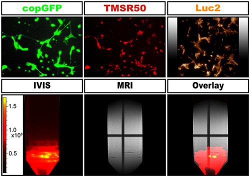
The application of stem cells in the treatment of various degenerative diseases is highly promising. However, cell‐based therapy could be limited by the problem of low viability of grafted cells and uncertainty about their fate. The combination of molecular imaging and contrast‐enhanced MRI may give more insight into the survival and behavior of grafted stem cells. We explore hair‐follicle‐bulge‐derived stem cells (HFBSCs) as a potential candidate for autologous cell‐based therapy. HFBSCs are transduced with a lentiviral construct with genes coding for bioluminescent (Luc2) and fluorescent (copGFP) reporter proteins, and subsequently loaded with magnetic nanoparticles to enable MRI visualization. Thus, we investigate for the first time if lentiviral transduction and cellular loading with nanoparticles have a cytotoxic effect upon these stem cells. Transduction efficiency, proliferation rate, cell viability and reporter protein co‐expression during long‐term culture of transduced HFBSCs were studied using fluorescence and bioluminescence microscopy. In addition, the effect of TMSR50 nanoparticles on proliferation and viability was investigated using the MTS assay and bioluminescence microscopy. The amount of TMSR50‐loaded HFBSCs needed to reach signal threshold for MRI was assessed using an agarose phantom. Transduction with the Luc2‐copGFP construct did not influence senescence, proliferation, doubling time, and differentiation of the HFBSCs. CopGFP expression was visible immediately after transduction and persisted for at least 15 passages, concomitantly with Luc2 expression. Cellular loading with TMSR50 nanoparticles did not affect cell viability and proliferation. The results imply that combined MRI and bioluminescence imaging may enable in vivo localization and long‐term monitoring of grafted viable HFBSCs. Copyright © 2016 John Wiley & Sons, Ltd.
中文翻译:

慢病毒转导和随后装载纳米颗粒不会影响毛囊隆起干细胞在体外的细胞活力和增殖
干细胞在各种退行性疾病的治疗中的应用是非常有前途的。但是,基于细胞的疗法可能会受到移植细胞生存力低和命运不确定的问题的限制。分子成像和对比增强MRI的结合可以为移植的干细胞的存活和行为提供更多的见识。我们探索毛囊隆突干细胞(HFBSC)作为基于自体细胞疗法的潜在候选者。用具有编码生物发光(Luc2)和荧光(copGFP)报告蛋白的基因的慢病毒构建体转导HFBSC,然后装载磁性纳米颗粒以进行MRI可视化。因此,我们首次研究慢病毒转导和纳米粒子对细胞的负载是否对这些干细胞具有细胞毒性作用。使用荧光和生物发光显微镜研究了转导的HFBSCs长期培养过程中的转导效率,增殖速率,细胞活力和报告蛋白共表达。此外,使用MTS分析和生物发光显微镜研究了TMSR50纳米颗粒对增殖和活力的影响。使用琼脂糖体模评估了达到MRI信号阈值所需的TMSR50加载的HFBSC的数量。Luc2-copGFP构建体的转导不影响HFBSC的衰老,增殖,倍增时间和分化。转导后立即可见CopGFP表达,并与Luc2表达同时持续至少15代。用TMSR50纳米颗粒加载细胞不会影响细胞活力和增殖。活的HFBSCs的体内定位和长期监测。版权所有©2016 John Wiley&Sons,Ltd.
更新日期:2016-12-15
中文翻译:
慢病毒转导和随后装载纳米颗粒不会影响毛囊隆起干细胞在体外的细胞活力和增殖
干细胞在各种退行性疾病的治疗中的应用是非常有前途的。但是,基于细胞的疗法可能会受到移植细胞生存力低和命运不确定的问题的限制。分子成像和对比增强MRI的结合可以为移植的干细胞的存活和行为提供更多的见识。我们探索毛囊隆突干细胞(HFBSC)作为基于自体细胞疗法的潜在候选者。用具有编码生物发光(Luc2)和荧光(copGFP)报告蛋白的基因的慢病毒构建体转导HFBSC,然后装载磁性纳米颗粒以进行MRI可视化。因此,我们首次研究慢病毒转导和纳米粒子对细胞的负载是否对这些干细胞具有细胞毒性作用。使用荧光和生物发光显微镜研究了转导的HFBSCs长期培养过程中的转导效率,增殖速率,细胞活力和报告蛋白共表达。此外,使用MTS分析和生物发光显微镜研究了TMSR50纳米颗粒对增殖和活力的影响。使用琼脂糖体模评估了达到MRI信号阈值所需的TMSR50加载的HFBSC的数量。Luc2-copGFP构建体的转导不影响HFBSC的衰老,增殖,倍增时间和分化。转导后立即可见CopGFP表达,并与Luc2表达同时持续至少15代。用TMSR50纳米颗粒加载细胞不会影响细胞活力和增殖。活的HFBSCs的体内定位和长期监测。版权所有©2016 John Wiley&Sons,Ltd.


 京公网安备 11010802027423号