当前位置:
X-MOL 学术
›
Contrast Media Mol. Imaging
›
论文详情
Our official English website, www.x-mol.net, welcomes your
feedback! (Note: you will need to create a separate account there.)
Surgical tissue handling methods to optimize ex vivo fluorescence with the activatable optical probe γ‐glutamyl hydroxymethyl rhodamine green
Contrast Media & Molecular Imaging Pub Date : 2016-07-22 , DOI: 10.1002/cmmi.1705 Toshiko Harada 1 , Yuko Nakamura 1 , Kazuhide Sato 1 , Tadanobu Nagaya 1 , Peter L. Choyke 1 , Yasuyuki Seto 2 , Hisataka Kobayashi 1
Contrast Media & Molecular Imaging Pub Date : 2016-07-22 , DOI: 10.1002/cmmi.1705 Toshiko Harada 1 , Yuko Nakamura 1 , Kazuhide Sato 1 , Tadanobu Nagaya 1 , Peter L. Choyke 1 , Yasuyuki Seto 2 , Hisataka Kobayashi 1
Affiliation
|
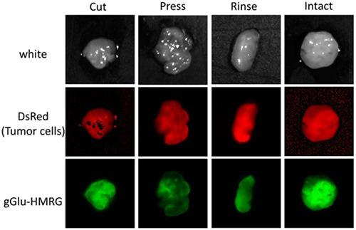
Optical fluorescence imaging has been developed as an aid to intraoperative diagnosis to improve surgical and endoscopic procedures. Compared with other intraoperative imaging methods, it is lower in cost, has a high safety margin, is portable and easy to use. γ‐glutamyl hydroxymethyl rhodamine green (gGlu‐HMRG) is a recently developed activatable fluorescence probe that emits strong fluorescence in the presence of the enzyme γ‐glutamyl transpeptidase (GGT), which is overexpressed in many cancers, including ovarian cancer. Ex vivo testing is important for clinical approval of such probes. The diagnostic performance of gGlu‐HMRG in fresh excised surgical specimens has been reported; however, details of tissue handling have not been optimized. In this study, we investigated four different tissue handling procedures to optimize imaging in excised tumor specimens. The fluorescence intensity time courses after the different tissue handling methods were compared. Additionally, the fluorescence positive areas were correlated with the presence of red fluorescent protein (RFP) in an RFP positive cell line as the standard of reference for cancer location. In the ‘intact’ groups, tumors yielded quick and homogeneous activation of gGlu‐HMRG. In the ‘rinse’ and ‘cut’ groups, the fluorescence intensity of the tumor was a little lower than that in the intact group. In the ‘pressed’ groups, however, fluorescence intensity from gGlu‐HMRG was lower over the entire time course, suggesting a decrease or relocation of excreted GGT. In conclusion, we demonstrate that the method of tissue handling prior to ex vivo imaging with the activatable probe gGlu‐HMRG has a strong influence on the signal derived from the specimen. Published 2016. This article is a U.S. Government work and is in the public domain in the USA.
中文翻译:

外科组织处理方法,使用可激活的光学探针γ-谷氨酰羟甲基若丹明绿优化体外荧光
已经开发了光学荧光成像作为术中诊断的辅助手段,以改善外科手术和内窥镜检查程序。与其他术中成像方法相比,它成本较低,安全系数高,便于携带且易于使用。γ-谷氨酰羟甲基罗丹明绿(gGlu-HMRG)是最近开发的一种可激活的荧光探针,在存在γ-谷氨酰转肽酶(GGT)的情况下会发出强荧光,该酶在许多癌症中都过度表达,包括卵巢癌。离体测试对于此类探头的临床批准很重要。已经报道了gGlu-HMRG在新鲜切除的手术标本中的诊断性能;但是,组织处理的细节尚未优化。在这项研究中,我们调查了四种不同的组织处理程序,以优化切除的肿瘤标本中的成像。比较了不同组织处理方法后的荧光强度时间进程。另外,荧光阳性区域与作为癌症定位参考标准的RFP阳性细胞系中红色荧光蛋白(RFP)的存在相关。在“完整”组中,肿瘤产生了快速且均一的gGlu-HMRG激活。在“冲洗”和“切割”组中,肿瘤的荧光强度略低于完整组。然而,在“受压”组中,gGlu-HMRG的荧光强度在整个时间过程中都较低,这表明排泄的GGT减少或重新定位。总之,我们证明了在处理之前的组织处理方法使用可激活探针gGlu-HMRG的离体成像对源自标本的信号有很大影响。2016年出版。本文是美国政府的工作,在美国属于公共领域。
更新日期:2016-07-22
中文翻译:
外科组织处理方法,使用可激活的光学探针γ-谷氨酰羟甲基若丹明绿优化体外荧光
已经开发了光学荧光成像作为术中诊断的辅助手段,以改善外科手术和内窥镜检查程序。与其他术中成像方法相比,它成本较低,安全系数高,便于携带且易于使用。γ-谷氨酰羟甲基罗丹明绿(gGlu-HMRG)是最近开发的一种可激活的荧光探针,在存在γ-谷氨酰转肽酶(GGT)的情况下会发出强荧光,该酶在许多癌症中都过度表达,包括卵巢癌。离体测试对于此类探头的临床批准很重要。已经报道了gGlu-HMRG在新鲜切除的手术标本中的诊断性能;但是,组织处理的细节尚未优化。在这项研究中,我们调查了四种不同的组织处理程序,以优化切除的肿瘤标本中的成像。比较了不同组织处理方法后的荧光强度时间进程。另外,荧光阳性区域与作为癌症定位参考标准的RFP阳性细胞系中红色荧光蛋白(RFP)的存在相关。在“完整”组中,肿瘤产生了快速且均一的gGlu-HMRG激活。在“冲洗”和“切割”组中,肿瘤的荧光强度略低于完整组。然而,在“受压”组中,gGlu-HMRG的荧光强度在整个时间过程中都较低,这表明排泄的GGT减少或重新定位。总之,我们证明了在处理之前的组织处理方法使用可激活探针gGlu-HMRG的离体成像对源自标本的信号有很大影响。2016年出版。本文是美国政府的工作,在美国属于公共领域。


 京公网安备 11010802027423号