Nature Communications ( IF 16.6 ) Pub Date : 2024-04-25 , DOI: 10.1038/s41467-024-47892-3 Quanyu Zhou , Chaim Glück , Lin Tang , Lukas Glandorf , Jeanne Droux , Mohamad El Amki , Susanne Wegener , Bruno Weber , Daniel Razansky , Zhenyue Chen
|
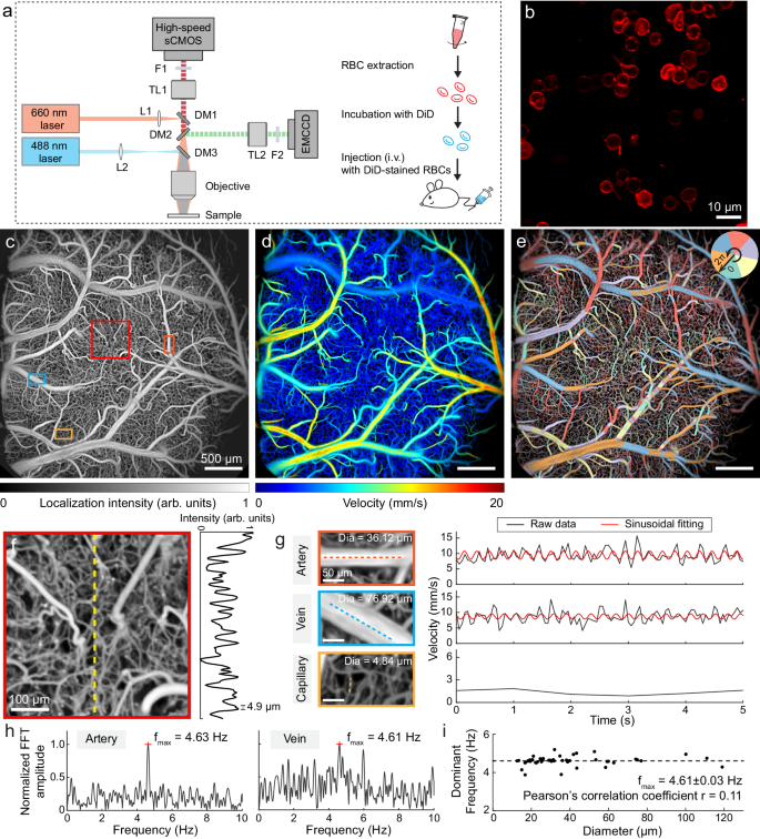
Large-scale imaging of brain activity with high spatio-temporal resolution is crucial for advancing our understanding of brain function. The existing neuroimaging techniques are largely limited by restricted field of view, slow imaging speed, or otherwise do not have the adequate spatial resolution to capture brain activities on a capillary and cellular level. To address these limitations, we introduce fluorescence localization microscopy aided with sparsely-labeled red blood cells for cortex-wide morphological and functional cerebral angiography with 4.9 µm spatial resolution and 1 s temporal resolution. When combined with fluorescence calcium imaging, the proposed method enables extended recordings of stimulus-evoked neuro-vascular changes in the murine brain while providing simultaneous multiparametric readings of intracellular neuronal activity, blood flow velocity/direction/volume, and vessel diameter. Owing to its simplicity and versatility, the proposed approach will become an invaluable tool for deciphering the regulation of cortical microcirculation and neurovascular coupling in health and disease.
中文翻译:
使用荧光标记的红细胞进行全皮质经颅定位显微镜
具有高时空分辨率的大脑活动的大规模成像对于增进我们对大脑功能的理解至关重要。现有的神经成像技术在很大程度上受到视野受限、成像速度慢或没有足够的空间分辨率来捕获毛细血管和细胞水平上的大脑活动的限制。为了解决这些限制,我们引入了荧光定位显微镜,辅助稀疏标记的红细胞,用于皮质范围的形态学和功能性脑血管造影,空间分辨率为 4.9 µm,时间分辨率为 1 s。当与荧光钙成像相结合时,所提出的方法能够延长记录小鼠大脑中刺激引起的神经血管变化,同时提供细胞内神经元活动、血流速度/方向/体积和血管直径的同步多参数读数。由于其简单性和多功能性,所提出的方法将成为破译健康和疾病中皮质微循环和神经血管耦合调节的宝贵工具。


 京公网安备 11010802027423号