当前位置:
X-MOL 学术
›
J. Diabetes Investig.
›
论文详情
Our official English website, www.x-mol.net, welcomes your feedback! (Note: you will need to create a separate account there.)
Characterization of the visually impaired patients with diabetes mellitus in Japan
Journal of Diabetes Investigation ( IF 3.2 ) Pub Date : 2024-03-27 , DOI: 10.1111/jdi.14195 Yuka Sugihara 1, 2 , Yoshihiro Takamura 1, 2 , Yutaka Yamada 2 , Masakazu Morioka 2 , Makoto Gozawa 2 , Kumiko Kato 1, 3 , Takao Hirano 1, 4 , Fumiko Murao 1, 5 , Miho Shimizu 1, 6 , Sentaro Kusuhara 1, 7 , Tomoya Murakami 1, 8 , Yuki Takenaka 1, 9 , Naoko Okabe 1, 10 , Tatsuya Jujo 1, 11 , Hiroto Terasaki 1, 12 , Daisuke Nagasato 1, 13 , Zhenyu Dong 14 , Shigeo Yoshida 1, 15 , Shuntaro Ogura 1, 16 , Kanako Yasuda 1, 17 , Gaku Ishigooka 1, 18 , Osamu Sawada 1, 19 , Fumiaki Higashijima 1, 20 , Masaru Inatani 2
Journal of Diabetes Investigation ( IF 3.2 ) Pub Date : 2024-03-27 , DOI: 10.1111/jdi.14195 Yuka Sugihara 1, 2 , Yoshihiro Takamura 1, 2 , Yutaka Yamada 2 , Masakazu Morioka 2 , Makoto Gozawa 2 , Kumiko Kato 1, 3 , Takao Hirano 1, 4 , Fumiko Murao 1, 5 , Miho Shimizu 1, 6 , Sentaro Kusuhara 1, 7 , Tomoya Murakami 1, 8 , Yuki Takenaka 1, 9 , Naoko Okabe 1, 10 , Tatsuya Jujo 1, 11 , Hiroto Terasaki 1, 12 , Daisuke Nagasato 1, 13 , Zhenyu Dong 14 , Shigeo Yoshida 1, 15 , Shuntaro Ogura 1, 16 , Kanako Yasuda 1, 17 , Gaku Ishigooka 1, 18 , Osamu Sawada 1, 19 , Fumiaki Higashijima 1, 20 , Masaru Inatani 2
Affiliation
|
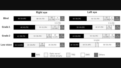
Aims/IntroductionTo conduct a multicenter survey of visually impaired patients with diabetes mellitus (DM) and to identify the physical and ocular characteristics that lead to blindness in Japan.Materials and MethodsVisually impaired patients with diabetes mellitus in Japan were divided into blind and low‐vision groups according to the World Health Organization classification. Data on parameters related to diabetes mellitus and ocular complications in the right and left eyes were collected from 19 highly advanced medical facilities and compared between the two groups.ResultsAmong 408 visually impaired persons (blind group: 257, low‐vision group: 151), 72.1% were under 70 years of age. The rates of neovascular glaucoma (NVG) (right eye, P = 0.041; left eye, P = 0.0031) or proliferative diabetic retinopathy (PDR) (right eye: P = 0.014, left eye: P = 0.0047) and the rate of proliferative membrane beyond half of the retinal area (right eye: P = 0.0263, left eye: P = 0.037) were significantly higher in the blind group. The direct cause of visual impairment was retinal atrophy, common in both groups. Neovascular glaucoma and diabetic macular edema were equally prevalent in the blind and low‐vision groups, respectively.ConclusionsIn Japan, blind patients with diabetes mellitus are characterized by severe conditions such as neovascular glaucoma and progressive proliferative diabetic retinopathy upon their initial visit to an advanced care facility. These results highlight the importance of monitoring retinopathy through regular ophthalmological examinations, internal medicine, and appropriate therapeutic intervention.
中文翻译:

日本糖尿病患者视力障碍的特征
目的/简介对日本糖尿病(DM)视力障碍患者进行多中心调查,并确定导致日本失明的身体和眼部特征。材料和方法日本糖尿病视力障碍患者分为失明和低视力根据世界卫生组织的分类。从19家高度先进的医疗机构收集了与糖尿病和左右眼眼部并发症相关的参数数据,并在两组之间进行比较。 结果在408名视力障碍者中(盲人组:257人,低视力组:151人), 72.1%的人年龄在70岁以下。新生血管性青光眼 (NVG) 的发病率(右眼、磷 = 0.041;左眼,磷 = 0.0031)或增殖性糖尿病视网膜病变(PDR)(右眼:磷 = 0.014,左眼:磷 = 0.0047)和超过一半视网膜区域的增殖膜率(右眼:磷 = 0.0263,左眼:磷 = 0.037)在盲组中显着较高。视力障碍的直接原因是视网膜萎缩,这在两组中都很常见。新生血管性青光眼和糖尿病性黄斑水肿在盲人和低视力群体中的发病率分别相同。结论在日本,患有糖尿病的盲人患者在初次就诊时会出现新生血管性青光眼和进行性增殖性糖尿病视网膜病变等严重病症。设施。这些结果强调了通过定期眼科检查、内科和适当的治疗干预来监测视网膜病变的重要性。
更新日期:2024-03-27
中文翻译:
日本糖尿病患者视力障碍的特征
目的/简介对日本糖尿病(DM)视力障碍患者进行多中心调查,并确定导致日本失明的身体和眼部特征。材料和方法日本糖尿病视力障碍患者分为失明和低视力根据世界卫生组织的分类。从19家高度先进的医疗机构收集了与糖尿病和左右眼眼部并发症相关的参数数据,并在两组之间进行比较。 结果在408名视力障碍者中(盲人组:257人,低视力组:151人), 72.1%的人年龄在70岁以下。新生血管性青光眼 (NVG) 的发病率(右眼、


 京公网安备 11010802027423号