Eye ( IF 3.9 ) Pub Date : 2024-03-21 , DOI: 10.1038/s41433-024-03021-4 Alaa E. Fayed , Martin J. Menten , Linus Kreitner , Johannes C. Paetzold , Daniel Rueckert , Sherry M. Bassily , Ramy R. Fikry , Ahmed M. Hagag , Sobha Sivaprasad
|
Objectives
To study the changes in vessel densities (VD) stratified by vessel diameter in the retinal superficial and deep vascular complexes (SVC/DVC) using optical coherence tomography angiography (OCTA) images obtained from people with diabetes and age-matched healthy controls.
Methods
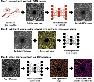
We quantified the VD based on vessel diameter categorized as <10, 10–20 and >20 μm in the SVC/DVC obtained on 3 × 3 mm2 OCTA scans using a deep learning-based segmentation and vascular graph extraction tool in people with diabetes and age-matched healthy controls.
Results
OCTA images obtained from 854 eyes of 854 subjects were divided into 5 groups: healthy controls (n = 555); people with diabetes with no diabetic retinopathy (DR, n = 90), mild and moderate non-proliferative DR (NPDR) (n = 96), severe NPDR (n = 42) and proliferative DR (PDR) (n = 71). Both SVC and DVC showed significant decrease in VD with increasing DR severity (p < 0.001). The largest difference was observed in the <10 μm vessels of the SVC between healthy controls and no DR (13.9% lower in no DR, p < 0.001). Progressive decrease in <10 μm vessels of the SVC and DVC was seen with increasing DR severity (p < 0.001). However, 10–20 μm vessels only showed decline in the DVC, but not the SVC (p < 0.001) and there was no change observed in the >20 μm vessels in either plexus.
Conclusions
Our findings suggest that OCTA is able to demonstrate a distinct vulnerability of the smallest retinal vessels in both plexuses that worsens with increasing severity of DR.
中文翻译:
不同直径和丛的视网膜脉管系统在不同严重程度的糖尿病视网膜病变中表现出明显的脆弱性
目标
使用从糖尿病患者和年龄匹配的健康对照者获得的光学相干断层扫描血管造影 (OCTA) 图像,研究视网膜浅层和深层血管复合体 (SVC/DVC) 中按血管直径分层的血管密度 (VD) 的变化。
方法
我们根据糖尿病患者使用基于深度学习的分割和血管图提取工具在 3 × 3 mm 2 OCTA 扫描中获得的 SVC/DVC 中的血管直径分类为 <10、10–20 和 >20 μm 来量化 VD和年龄匹配的健康对照。
结果
从 854 名受试者的 854 只眼睛获得的 OCTA 图像分为 5 组:健康对照组 ( n = 555);无糖尿病视网膜病变的糖尿病患者 (DR, n = 90)、轻度和中度非增殖性 DR (NPDR) ( n = 96)、重度 NPDR ( n = 42) 和增殖性 DR (PDR) ( n = 71)。随着 DR 严重程度的增加,SVC 和 DVC 的 VD 均显着降低 ( p < 0.001)。健康对照和无 DR 之间的 SVC <10 μm 血管中观察到的最大差异(无 DR 中低 13.9%,p < 0.001)。随着 DR 严重程度的增加,SVC 和 DVC <10 μm 血管逐渐减少 ( p < 0.001)。然而,10-20 μm 血管仅显示 DVC 下降,而 SVC 没有下降 ( p < 0.001),并且任一丛中 >20 μm 血管均未观察到变化。
结论
我们的研究结果表明,OCTA 能够显示两个神经丛中最小视网膜血管的明显脆弱性,这种脆弱性随着 DR 严重程度的增加而恶化。


 京公网安备 11010802027423号