Surgical Endoscopy ( IF 3.1 ) Pub Date : 2024-03-11 , DOI: 10.1007/s00464-024-10758-2 Nazanin Safavian , Simon K. C. Toh , Martino Pani , Raymond Lee
|
Background
Accurate measurement of polyps size is crucial in predicting malignancy, planning relevant intervention strategies and surveillance schedules. Endoscopists’ visual estimations can lack precision. This study builds on our prior research, with the aim to evaluate a recently developed quantitative method to measure the polyp size and location accurately during a simulated endoscopy session.
Methods
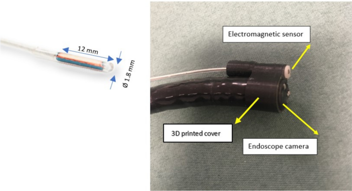
The quantitative method merges information about endoscopic positions obtained from an electromagnetic tracking sensor, with corresponding points on the images of the segmented polyp border. This yields real-scale 3D coordinates of the border of the polyp. By utilising the sensor, positions of any anatomical landmarks are attainable, enabling the estimation of a polyp’s location relative to them. To verify the method’s reliability and accuracy, simulated endoscopies were conducted in pig stomachs, where polyps were artificially created and assessed in a test–retest manner. The polyp measurements were subsequently compared against clipper measurements.
Results
The average size of the fifteen polyps evaluated was approximately 12 ± 4.3 mm, ranging from 5 to 20 mm. The test–retest reliability, measured by the Intraclass Correlation Coefficient (ICC) for polyp size estimation, demonstrated an absolute agreement of 0.991 (95% CI 0.973–0.997, p < 0.05). Bland & Altman analysis revealed a mean estimation difference of − 0.17 mm (− 2.03%) for polyp size and, a mean difference of − 0.4 mm (− 0.21%) for polyp location. Both differences were statistically non-significant (p > 0.05). When comparing the proposed method with calliper measurements, the Bland & Altman plots showed 95% of size estimation differences between − 1.4 and 1.8 mm (− 13 to 17.4%) which was not significant (p > 0.05).
Conclusions
The proposed method of measurements of polyp size and location was found to be highly accurate, offering great potential for clinical implementation to improve polyp assessment. This level of performance represents a notable improvement over visual estimation technique used in clinical practice.
中文翻译:
增强内窥镜测量:验证上消化道内窥镜检查中息肉大小和位置估计的定量方法
背景
准确测量息肉大小对于预测恶性肿瘤、规划相关干预策略和监测计划至关重要。内窥镜医师的视觉估计可能不够精确。这项研究建立在我们之前的研究基础上,旨在评估最近开发的定量方法,以在模拟内窥镜检查过程中准确测量息肉的大小和位置。
方法
该定量方法将从电磁跟踪传感器获得的内窥镜位置信息与分割的息肉边界图像上的对应点合并。这会产生息肉边界的真实比例 3D 坐标。通过利用传感器,可以获得任何解剖标志的位置,从而能够估计息肉相对于它们的位置。为了验证该方法的可靠性和准确性,在猪胃中进行了模拟内窥镜检查,人工产生息肉并以重测的方式进行评估。随后将息肉测量值与剪子测量值进行比较。
结果
所评估的 15 个息肉的平均大小约为 12 ± 4.3 毫米,范围为 5 至 20 毫米。通过息肉大小估计的组内相关系数 (ICC) 测量的重测可靠性显示,绝对一致性为 0.991(95% CI 0.973–0.997,p < 0.05)。Bland & Altman 分析显示,息肉大小的平均估计差异为 − 0.17 毫米(− 2.03%),息肉位置的平均估计差异为 − 0.4 毫米(− 0.21%)。两种差异在统计学上均不显着(p > 0.05)。将所提出的方法与卡尺测量进行比较时,Bland & Altman 图显示 95% 的尺寸估计差异在 − 1.4 和 1.8 mm 之间(− 13 到 17.4%),但并不显着(p > 0.05)。
结论
研究发现,所提出的测量息肉大小和位置的方法非常准确,为临床实施改善息肉评估提供了巨大的潜力。这种性能水平代表了临床实践中使用的视觉估计技术的显着改进。


 京公网安备 11010802027423号