Surgical Endoscopy ( IF 3.1 ) Pub Date : 2024-03-08 , DOI: 10.1007/s00464-023-10615-8 Anas Amin Preukschas , Philipp Anthony Wise , Lisa Bettscheider , Micha Pfeiffer , Martin Wagner , Matthias Huber , Mohammad Golriz , Lars Fischer , Arianeb Mehrabi , Fabian Rössler , Stefanie Speidel , Thilo Hackert , Beat Peter Müller-Stich , Felix Nickel , Hannes Götz Kenngott
|
Objective
Evaluation of the benefits of a virtual reality (VR) environment with a head-mounted display (HMD) for decision-making in liver surgery.
Background
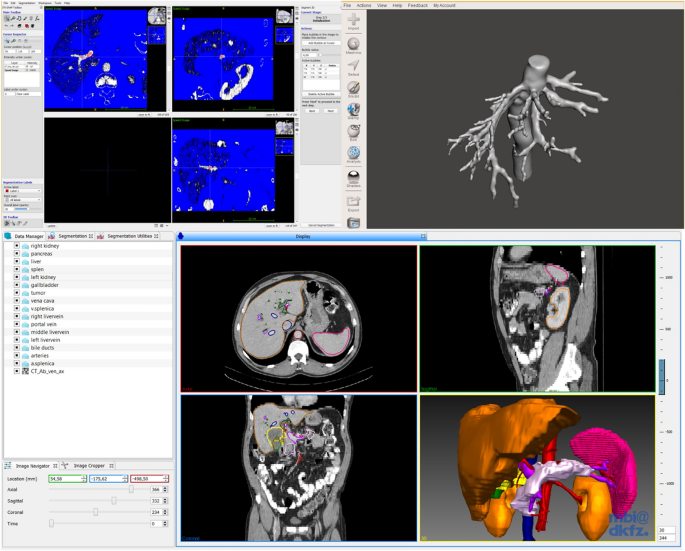
Training in liver surgery involves appraising radiologic images and considering the patient’s clinical information. Accurate assessment of 2D-tomography images is complex and requires considerable experience, and often the images are divorced from the clinical information. We present a comprehensive and interactive tool for visualizing operation planning data in a VR environment using a head-mounted-display and compare it to 3D visualization and 2D-tomography.
Methods
Ninety medical students were randomized into three groups (1:1:1 ratio). All participants analyzed three liver surgery patient cases with increasing difficulty. The cases were analyzed using 2D-tomography data (group “2D”), a 3D visualization on a 2D display (group “3D”) or within a VR environment (group “VR”). The VR environment was displayed using the “Oculus Rift ™” HMD technology. Participants answered 11 questions on anatomy, tumor involvement and surgical decision-making and 18 evaluative questions (Likert scale).
Results
Sum of correct answers were significantly higher in the 3D (7.1 ± 1.4, p < 0.001) and VR (7.1 ± 1.4, p < 0.001) groups than the 2D group (5.4 ± 1.4) while there was no difference between 3D and VR (p = 0.987). Times to answer in the 3D (6:44 ± 02:22 min, p < 0.001) and VR (6:24 ± 02:43 min, p < 0.001) groups were significantly faster than the 2D group (09:13 ± 03:10 min) while there was no difference between 3D and VR (p = 0.419). The VR environment was evaluated as most useful for identification of anatomic anomalies, risk and target structures and for the transfer of anatomical and pathological information to the intraoperative situation in the questionnaire.
Conclusions
A VR environment with 3D visualization using a HMD is useful as a surgical training tool to accurately and quickly determine liver anatomy and tumor involvement in surgery.
中文翻译:
比较虚拟现实头戴式显示器与屏幕上的三维可视化和二维计算机断层扫描数据,用于肝外科决策培训:一项随机对照研究
客观的
评估带有头戴式显示器 (HMD) 的虚拟现实 (VR) 环境对于肝脏手术决策的益处。
背景
肝脏手术培训包括评估放射图像并考虑患者的临床信息。二维断层扫描图像的准确评估非常复杂,需要大量的经验,而且图像通常与临床信息脱节。我们提出了一种全面的交互式工具,用于使用头戴式显示器在 VR 环境中可视化操作规划数据,并将其与 3D 可视化和 2D 断层扫描进行比较。
方法
90 名医学生被随机分为三组(比例为 1:1:1)。所有参与者分析了三个难度逐渐增加的肝脏手术患者病例。使用 2D 断层扫描数据(“2D”组)、2D 显示器上的 3D 可视化(“3D”组)或 VR 环境(“VR”组)对病例进行分析。VR环境使用“Oculus Rift™”HMD技术来显示。参与者回答了 11 个有关解剖学、肿瘤累及和手术决策的问题以及 18 个评估问题(李克特量表)。
结果
3D(7.1 ± 1.4,p < 0.001)和 VR(7.1 ± 1.4,p < 0.001)组的正确答案总和显着高于 2D 组(5.4 ± 1.4),而 3D 和 VR 之间没有差异(p = 0.987)。3D(6:44 ± 02:22 分钟,p < 0.001)和 VR(6:24 ± 02:43 分钟,p < 0.001)组的回答时间明显快于 2D 组(09:13 ± 03) :10 分钟),而 3D 和 VR 之间没有差异(p = 0.419)。VR 环境被认为对于识别解剖异常、风险和目标结构以及将解剖和病理信息传输到问卷中的术中情况最有用。
结论
使用 HMD 进行 3D 可视化的 VR 环境可用作手术训练工具,可准确快速地确定手术中的肝脏解剖结构和肿瘤参与情况。


 京公网安备 11010802027423号