Brain Imaging and Behavior ( IF 3.2 ) Pub Date : 2024-02-26 , DOI: 10.1007/s11682-024-00866-x Hong-Yu Lin , Hui-Wei Huang , Qiu-Yi Dong , Li-Min Cai , Hua-Jun Chen
|
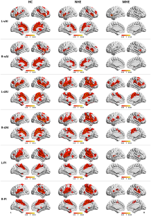
We investigated abnormal functional connectivity (FC) patterns of insular subregions in patients with minimal hepatic encephalopathy (MHE) and examined their relationships with cognitive dysfunction using resting-state functional magnetic resonance imaging (fMRI). We collected resting-state fMRI data in 54 patients with cirrhosis [20 with MHE and 34 without MHE (NHE)] and 25 healthy controls. After defining six subregions of insula, we mapped whole-brain FC of the insular subregions and identified FC differences through three groups. FC of the insular subregions was correlated against clinical parameters (including venous blood ammonia level, Child-Pugh score, and cognitive score). The discrimination performance between the MHE and NHE groups was evaluated by performing a classification analysis using the FC index. Across three groups, the observed FC differences involved four insular subregions, including the left-ventral anterior insula, left-dorsal anterior insula, right-dorsal anterior insula, and left-posterior insula (P < 0.05 with false discovery rate correction). Moreover, the FC of these four insular subregions progressively attenuated from NHE to MHE. In addition, hypoconnectivity of insular subregions was correlated with the poor neuropsychological performance and the evaluated blood ammonia levels in patients (P < 0.05 with Bonferroni correction). The FC of insular subregions yielded moderate discriminative value between the MHE and NHE groups (AUC = 0.696–0.809). FC disruption of insular subregions is related to worse cognitive performance in MHE. This study extended our understanding about the neurophysiology of MHE and may assist for its diagnosis.
中文翻译:
轻度肝性脑病肝硬化患者岛叶亚区功能连接破坏
我们研究了轻微肝性脑病(MHE)患者岛叶亚区域的异常功能连接(FC)模式,并使用静息态功能磁共振成像(fMRI)检查了它们与认知功能障碍的关系。我们收集了 54 名肝硬化患者(20 名患有 MHE,34 名不患有 MHE (NHE))和 25 名健康对照者的静息态 fMRI 数据。在定义了岛叶的六个子区域后,我们绘制了岛叶子区域的全脑 FC,并通过三组确定了 FC 差异。岛叶次区域的 FC 与临床参数(包括静脉血氨水平、Child-Pugh 评分和认知评分)相关。通过使用 FC 指数进行分类分析来评估 MHE 和 NHE 组之间的辨别性能。在三组中,观察到的 FC 差异涉及四个岛叶分区,包括左腹前岛叶、左背前岛叶、右背前岛叶和左后岛叶(P < 0.05,错误发现率校正)。此外,这四个岛状分区的FC从NHE到MHE逐渐衰减。此外,岛叶亚区连接性低下与患者较差的神经心理学表现和评估的血氨水平相关(经Bonferroni校正后P < 0.05)。岛叶次区域的 FC 在 MHE 和 NHE 组之间产生中等的区分价值 (AUC = 0.696–0.809)。岛叶次区域的 FC 破坏与 MHE 认知表现较差有关。这项研究扩展了我们对 MHE 神经生理学的理解,并可能有助于其诊断。


 京公网安备 11010802027423号