IRBM ( IF 4.8 ) Pub Date : 2023-12-27 , DOI: 10.1016/j.irbm.2023.100818 David Lemonnier , Ikram Mezghani , Georgios Theocharidis , Brandon J. Sumpio , Samuel K. Sia , Aristidis Veves , Parag V. Chitnis
|
Background
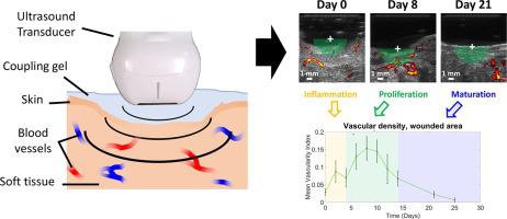
Monitoring of wound healing progression is critical due to the risk of infection, non-healing wounds, or evolution towards a chronic state. Tissue vasculature is one of the most representative features reflecting healing status. This study explores the feasibility of vascular ultrasound imaging of open wounds and the extraction of vascular-related features in a longitudinal study.
Material and methods
C57BL/6 mice received a 1 cm-diameter full-thickness wound on their dorsum and were imaged using ultrasound from the surgical day (Day 0) to 25 days post-wounding. The high frame rate, plane waves acquisitions with a 15 MHz transducer were postprocessed with Singular Value Decomposition (SVD) filtering to provide vascular information.
Results
Vascularity Index (VI) calculations showed an increased vascular signal in the wound from Day 2 post-wounding and were significantly higher from day 6 to day 10 post-wounding compared to Day 0 (p<0.05). VI values were back to the basal level after 3 weeks. In comparison, no significant difference was highlighted for the vascular signal in the peri-wound area.
Conclusions
These results show that vascular ultrasound imaging can be applied to track vascular changes of open wounds during the healing process. This approach may also be extended to other types of wounds for detecting early signs likely to cause complications.
中文翻译:
用于评估伤口愈合过程中血管重塑的无对比高帧率超声成像
背景
由于存在感染、伤口不愈合或演变为慢性状态的风险,监测伤口愈合进展至关重要。组织脉管系统是反映愈合状态的最具代表性的特征之一。本研究探讨了在纵向研究中对开放性伤口进行血管超声成像并提取血管相关特征的可行性。
材料与方法
C57BL/6 小鼠的背部受到直径 1 厘米的全层伤口,并从手术当天(第 0 天)到受伤后 25 天使用超声波进行成像。使用 15 MHz 传感器采集的高帧速率平面波经过奇异值分解 (SVD) 滤波进行后处理,以提供血管信息。
结果
血管指数 (VI) 计算显示,伤口中的血管信号从受伤后第 2 天开始增加,并且与第 0 天相比,从受伤后第 6 天到第 10 天显着升高 (p<0.05)。3周后VI值恢复到基础水平。相比之下,伤口周围区域的血管信号没有显着差异。
结论
这些结果表明,血管超声成像可用于跟踪开放性伤口愈合过程中的血管变化。这种方法也可以扩展到其他类型的伤口,以检测可能导致并发症的早期迹象。


 京公网安备 11010802027423号