当前位置:
X-MOL 学术
›
Ann. N. Y. Acad. Sci.
›
论文详情
Our official English website, www.x-mol.net, welcomes your feedback! (Note: you will need to create a separate account there.)
Segregation of neuronal and vascular retinal damage in patients with hypertension and diabetes
Annals of the New York Academy of Sciences ( IF 5.2 ) Pub Date : 2023-12-12 , DOI: 10.1111/nyas.15089 Jacqueline Chua 1, 2 , Damon Wong 1, 3, 4 , Ai Ping Yow 1, 3, 5, 6 , Bingyao Tan 1, 3, 5 , Xinyu Liu 1, 2, 3 , Munirah Binte Ismail 1, 3, 5 , Calvin Woon Loong Chin 2, 7 , Ecosse Lamoureux 1, 2 , Rahat Husain 1, 2 , Leopold Schmetterer 1, 2, 3, 4, 5
Annals of the New York Academy of Sciences ( IF 5.2 ) Pub Date : 2023-12-12 , DOI: 10.1111/nyas.15089 Jacqueline Chua 1, 2 , Damon Wong 1, 3, 4 , Ai Ping Yow 1, 3, 5, 6 , Bingyao Tan 1, 3, 5 , Xinyu Liu 1, 2, 3 , Munirah Binte Ismail 1, 3, 5 , Calvin Woon Loong Chin 2, 7 , Ecosse Lamoureux 1, 2 , Rahat Husain 1, 2 , Leopold Schmetterer 1, 2, 3, 4, 5
Affiliation
|
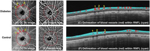
This study aimed to examine the impact of diabetes and hypertension on retinal nerve fiber layer (RNFL) thickness components. Optical coherence tomography (OCT) measurements do not consider blood vessel contribution, which this study addressed. We hypothesized that diabetes and/or hypertension would lead to thinner RNFL versus controls due to the vascular component. OCT angiography was used to measure the RNFL in 121 controls, 50 diabetes patients, 371 hypertension patients, and 177 diabetes patients with hypertension. A novel technique separated the RNFL thickness into original (vascular component) and corrected (no vascular component) measurements. Diabetes-only (98 ± 1.7 µm; p = 0.002) and diabetes with hypertension (99 ± 0.8 µm; p = 0.001) patients had thinner original RNFL versus controls (102 ± 0.8 µm). No difference was seen between hypertension-only patients (101 ± 0.5 µm; p = 0.083) and controls. After removing the blood vessel component, diabetes/hypertension groups had thinner corrected RNFL versus controls (p = 0.024). Discrepancies in diabetes/hypertension patients were due to thicker retinal blood vessels within the RNFL thickness (p = 0.002). Our findings suggest that diabetes and/or hypertension independently contribute to neurodegenerative thinning of the RNFL, even in the absence of retinopathy. The differentiation of neuronal and vascular components in RNFL thickness measurements provided by the novel technique highlights the importance of considering vascular changes in individuals with these conditions.
中文翻译:

高血压和糖尿病患者神经元和血管视网膜损伤的分离
本研究旨在探讨糖尿病和高血压对视网膜神经纤维层 (RNFL) 厚度成分的影响。光学相干断层扫描 (OCT) 测量不考虑血管的影响,而本研究正是解决了这一问题。我们假设,由于血管成分,糖尿病和/或高血压会导致 RNFL 较对照组更薄。采用 OCT 血管造影测量 121 名对照者、50 名糖尿病患者、371 名高血压患者和 177 名糖尿病合并高血压患者的 RNFL。一项新技术将 RNFL 厚度分为原始(血管成分)和校正(无血管成分)测量值。与对照组 (102 ± 0.8 µm) 相比,单纯糖尿病 (98 ± 1.7 µm; p = 0.002) 和糖尿病合并高血压 (99 ± 0.8 µm; p = 0.001) 患者的原始 RNFL 较薄。仅高血压患者 (101 ± 0.5 µm; p = 0.083) 和对照之间没有发现差异。去除血管成分后,糖尿病/高血压组的校正 RNFL 较对照组更薄(p = 0.024)。糖尿病/高血压患者的差异是由于 RNFL 厚度内的视网膜血管较厚 ( p = 0.002)。我们的研究结果表明,即使在没有视网膜病变的情况下,糖尿病和/或高血压也会独立导致 RNFL 的神经退行性变薄。新技术提供的 RNFL 厚度测量中神经元和血管成分的区分凸显了考虑患有这些疾病的个体血管变化的重要性。
更新日期:2023-12-12
中文翻译:
高血压和糖尿病患者神经元和血管视网膜损伤的分离
本研究旨在探讨糖尿病和高血压对视网膜神经纤维层 (RNFL) 厚度成分的影响。光学相干断层扫描 (OCT) 测量不考虑血管的影响,而本研究正是解决了这一问题。我们假设,由于血管成分,糖尿病和/或高血压会导致 RNFL 较对照组更薄。采用 OCT 血管造影测量 121 名对照者、50 名糖尿病患者、371 名高血压患者和 177 名糖尿病合并高血压患者的 RNFL。一项新技术将 RNFL 厚度分为原始(血管成分)和校正(无血管成分)测量值。与对照组 (102 ± 0.8 µm) 相比,单纯糖尿病 (98 ± 1.7 µm; p = 0.002) 和糖尿病合并高血压 (99 ± 0.8 µm; p = 0.001) 患者的原始 RNFL 较薄。仅高血压患者 (101 ± 0.5 µm; p = 0.083) 和对照之间没有发现差异。去除血管成分后,糖尿病/高血压组的校正 RNFL 较对照组更薄(p = 0.024)。糖尿病/高血压患者的差异是由于 RNFL 厚度内的视网膜血管较厚 ( p = 0.002)。我们的研究结果表明,即使在没有视网膜病变的情况下,糖尿病和/或高血压也会独立导致 RNFL 的神经退行性变薄。新技术提供的 RNFL 厚度测量中神经元和血管成分的区分凸显了考虑患有这些疾病的个体血管变化的重要性。


 京公网安备 11010802027423号