当前位置:
X-MOL 学术
›
Ann. Noninvasive Electrocardiol.
›
论文详情
Our official English website, www.x-mol.net, welcomes your feedback! (Note: you will need to create a separate account there.)
ISE/ISHNE expert consensus statement on the ECG diagnosis of left ventricular hypertrophy: The change of the paradigm
Annals of Noninvasive Electrocardiology ( IF 1.9 ) Pub Date : 2023-11-24 , DOI: 10.1111/anec.13097 Ljuba Bacharova 1 , Philippe Chevalier 2, 3 , Bulent Gorenek 4 , Christian Jons 5 , Yi-Gang Li 6 , Emanuela T Locati 7 , Maren Maanja 8 , Andrés Ricardo Pérez-Riera 9, 10 , Pyotr G Platonov 11 , Antonio Luiz Pinho Ribeiro 12, 13 , Douglas Schocken 14 , Elsayed Z Soliman 15 , Jana Svehlikova 16 , Larisa G Tereshchenko 17 , Martin Ugander 18, 19 , Niraj Varma 20 , Zaklyazminskaya Elena 21 , Takanori Ikeda 22
Annals of Noninvasive Electrocardiology ( IF 1.9 ) Pub Date : 2023-11-24 , DOI: 10.1111/anec.13097 Ljuba Bacharova 1 , Philippe Chevalier 2, 3 , Bulent Gorenek 4 , Christian Jons 5 , Yi-Gang Li 6 , Emanuela T Locati 7 , Maren Maanja 8 , Andrés Ricardo Pérez-Riera 9, 10 , Pyotr G Platonov 11 , Antonio Luiz Pinho Ribeiro 12, 13 , Douglas Schocken 14 , Elsayed Z Soliman 15 , Jana Svehlikova 16 , Larisa G Tereshchenko 17 , Martin Ugander 18, 19 , Niraj Varma 20 , Zaklyazminskaya Elena 21 , Takanori Ikeda 22
Affiliation
|
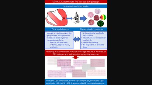
The ECG diagnosis of LVH is predominantly based on the QRS voltage criteria. The classical paradigm postulates that the increased left ventricular mass generates a stronger electrical field, increasing the leftward and posterior QRS forces, reflected in the augmented QRS amplitude. However, the low sensitivity of voltage criteria has been repeatedly documented. We discuss possible reasons for this shortcoming and proposal of a new paradigm. The theoretical background for voltage measured at the body surface is defined by the solid angle theorem, which relates the measured voltage to spatial and non-spatial determinants. The spatial determinants are represented by the extent of the activation front and the distance of the recording electrodes. The non-spatial determinants comprise electrical characteristics of the myocardium, which are comparatively neglected in the interpretation of the QRS patterns. Various clinical conditions are associated with LVH. These conditions produce considerable diversity of electrical properties alterations thereby modifying the resultant QRS patterns. The spectrum of QRS patterns observed in LVH patients is quite broad, including also left axis deviation, left anterior fascicular block, incomplete and complete left bundle branch blocks, Q waves, and fragmented QRS. Importantly, the QRS complex can be within normal limits. The new paradigm stresses the electrophysiological background in interpreting QRS changes, i.e., the effect of the non-spatial determinants. This postulates that the role of ECG is not to estimate LV size in LVH, but to understand and decode the underlying electrical processes, which are crucial in relation to cardiovascular risk assessment.
中文翻译:

ISE/ISHNE 关于左心室肥厚心电图诊断的专家共识声明:范式的改变
LVH 的心电图诊断主要基于 QRS 电压标准。经典范式假设增加的左心室质量会产生更强的电场,从而增加向左和后向的 QRS 波力量,这反映在 QRS 波幅的增强上。然而,电压标准的低灵敏度已被多次记录。我们讨论了这一缺点的可能原因并提出了新范式。在身体表面测量的电压的理论背景由立体角定理定义,该定理将测量的电压与空间和非空间决定因素联系起来。空间决定因素由激活前沿的范围和记录电极的距离表示。非空间决定因素包括心肌的电特性,这些特性在 QRS 模式的解释中相对被忽略。多种临床状况与 LVH 相关。这些条件会产生相当多的电特性变化,从而改变生成的 QRS 模式。在 LVH 患者中观察到的 QRS 波谱非常广泛,还包括电轴左偏、左前分支传导阻滞、不完全和完全左束支传导阻滞、Q 波和碎片 QRS。重要的是,QRS 波群可以在正常范围内。新范式强调解释 QRS 变化的电生理学背景,即非空间决定因素的影响。这假设心电图的作用不是估计 LVH 中的 LV 大小,而是理解和解码潜在的电过程,这对于心血管风险评估至关重要。
更新日期:2023-11-24
中文翻译:
ISE/ISHNE 关于左心室肥厚心电图诊断的专家共识声明:范式的改变
LVH 的心电图诊断主要基于 QRS 电压标准。经典范式假设增加的左心室质量会产生更强的电场,从而增加向左和后向的 QRS 波力量,这反映在 QRS 波幅的增强上。然而,电压标准的低灵敏度已被多次记录。我们讨论了这一缺点的可能原因并提出了新范式。在身体表面测量的电压的理论背景由立体角定理定义,该定理将测量的电压与空间和非空间决定因素联系起来。空间决定因素由激活前沿的范围和记录电极的距离表示。非空间决定因素包括心肌的电特性,这些特性在 QRS 模式的解释中相对被忽略。多种临床状况与 LVH 相关。这些条件会产生相当多的电特性变化,从而改变生成的 QRS 模式。在 LVH 患者中观察到的 QRS 波谱非常广泛,还包括电轴左偏、左前分支传导阻滞、不完全和完全左束支传导阻滞、Q 波和碎片 QRS。重要的是,QRS 波群可以在正常范围内。新范式强调解释 QRS 变化的电生理学背景,即非空间决定因素的影响。这假设心电图的作用不是估计 LVH 中的 LV 大小,而是理解和解码潜在的电过程,这对于心血管风险评估至关重要。


 京公网安备 11010802027423号