Lung ( IF 5 ) Pub Date : 2023-11-14 , DOI: 10.1007/s00408-023-00655-1 Marc A Judson 1 , Jianwei Qiu 2 , Camille L Dumas 3 , Jun Yang 4 , Brion Sarachan 2 , Jhimli Mitra 2
|
Purpose
To determine the reliability of an artificial intelligence, deep learning (AI/DL)-based method of chest computer tomography (CT) scan analysis to distinguish pulmonary sarcoidosis from negative lung cancer screening chest CT scans (Lung Imaging Reporting and Data System score 1, Lung-RADS score 1).
Methods
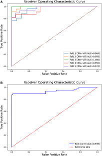
Chest CT scans of pulmonary sarcoidosis were evaluated by a clinician experienced with sarcoidosis and a chest radiologist for clinical and radiologic evidence of sarcoidosis and exclusion of alternative or concomitant pulmonary diseases. The AI/DL based method used an ensemble network architecture combining Convolutional Neural Networks (CNNs) and Vision Transformers (ViTs). The method was applied to 126 pulmonary sarcoidosis and 96 Lung-RADS score 1 CT scans. The analytic approach of training and validation of the AI/DL method used a fivefold cross-validation technique, where 4/5th of the available data set was used to train a diagnostic model and tested on the remaining 1/5th of the data set, and repeated 4 more times with non-overlapping validation/test data. The probability values were used to generate Receiver Operating Characteristic (ROC) curves to assess the model’s discriminatory power.
Results
The sensitivity, specificity, positive and negative predictive value of the AI/DL method for the 5 folds of the training/validation sets and the entire set of CT scans were all over 94% to distinguish pulmonary sarcoidosis from LUNG-RADS score 1 chest CT scans. The area under the curve for the corresponding ROC curves were all over 97%.
Conclusion
This AL/DL model shows promise to distinguish sarcoidosis from alternative pulmonary conditions using minimal radiologic data.
中文翻译:
用于肺结节病放射诊断的人工智能平台:胸部计算机断层扫描分析的初步初步研究,以区分肺结节病和阴性肺癌筛查扫描
目的
确定基于人工智能、深度学习 (AI/DL) 的胸部计算机断层扫描 (CT) 扫描分析方法的可靠性,以区分肺结节病和阴性肺癌筛查胸部 CT 扫描(肺部 影像报告和数据系统评分 1,肺-RADS 评分 1)。
方法
肺结节病的胸部 CT 扫描由具有结节病经验的临床医生和胸部放射科医生进行评估,以获取结节病的临床和放射学证据,并排除其他或伴随的肺部疾病。基于 AI/DL 的方法使用结合了卷积神经网络 (CNN) 和视觉变换器 (ViT) 的集成网络架构。该方法适用于126例肺结节病和96例Lung-RADS评分1的CT扫描。AI/DL方法的训练和验证的分析方法使用五重交叉验证技术,其中4/5的可用数据集用于训练诊断模型并在剩余的1/5数据集上进行测试,并使用不重叠的验证/测试数据重复 4 次。概率值用于生成受试者工作特征 (ROC) 曲线,以评估模型的辨别力。
结果
AI/DL方法对5倍训练/验证集和整组CT扫描的敏感性、特异性、阳性和阴性预测值均超过94%,以区分肺结节病和LUNG-RADS评分1胸部CT扫描。相应的ROC曲线的曲线下面积均超过97%。
结论
该 AL/DL 模型有望利用最少的放射学数据区分结节病和其他肺部疾病。


 京公网安备 11010802027423号