Annals of Nuclear Medicine ( IF 2.6 ) Pub Date : 2023-10-03 , DOI: 10.1007/s12149-023-01871-8 Cheng Liu 1, 2, 3, 4, 5 , Guang Ma 4 , Jiangang Zhang 2, 3, 6 , Jingyi Cheng 2, 3, 6 , Zhongyi Yang 4 , Shaoli Song 1, 2, 3, 4, 5
|
Objective
The aim of this study was to investigate the potential value of dual tracers 18F-FDG and 18F-FES PET/CT in predicting response to Cyclin-Dependent 4/6 Kinase (CDK4/6) inhibitors combined with endocrine therapy for metastatic estrogen receptor (ER)-positive breast cancer patients.
Methods
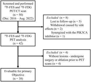
This retrospective study enrolled 38 ER-positive metastatic breast cancer patients from our center who underwent both 18F-FDG and 18F-FES PET/CT scans within 1 month before CDK4/6 inhibitors combined with endocrine therapy. The extracted parameters comprised the maximum standardized uptake value (SUVmax) for both FDG and FES PET, as well as the ratio between FES and FDG SUVmax. Each parameter was dichotomized based on its median threshold. The primary endpoint was progression-free survival (PFS), which was estimated using the Kaplan–Meier method and compared by the log-rank test.
Results
After a median follow-up of 15.6 months, progressive disease was observed in 23 out of 38 patients, and the median PFS for the whole cohort was 21.0 months [95% confidence interval (CI) 12.7–29.3]. FES and FDG PET identified 6 patients (15.8%) with FES-negative lesions, suggesting ER heterogeneity in metastatic lesions. The median PFS of these patients was only 5.3 months (95% CI 1.7–8.9), which was substantially shorter than that of patients with 100% FES-positive lesions (median PFS 22.9 months, 95% CI 17.1–28.7, P < 0.001). Patients with 100% FES-positive lesions who had high FES/FDG showed significantly shorter PFS compared to those with low FES/FDG (14.9 vs. 30.5 months, P = 0.003).
Conclusions
This study shows that FDG and FES PET imaging may serve as valuable tools for patient selection in the context of CDK4/6 inhibitor therapy combined with endocrine treatment, and have the potential to function as prognostic biomarkers.
中文翻译:
18F-FES 和 18F-FDG PET/CT 成像作为接受细胞周期蛋白依赖性 4/6 激酶抑制剂联合内分泌治疗的转移性乳腺癌患者的预测生物标志物
客观的
本研究的目的是探讨双示踪剂18 F-FDG 和18 F-FES PET/CT 在预测细胞周期蛋白依赖性 4/6 激酶 (CDK4/6) 抑制剂联合内分泌治疗转移性雌激素疗效中的潜在价值受体(ER)阳性乳腺癌患者。
方法
本回顾性研究纳入了我中心38例ER阳性转移性乳腺癌患者,在CDK4/6抑制剂联合内分泌治疗前1个月内接受18F -FDG和18F -FES PET/CT扫描。提取的参数包括 FDG 和 FES PET 的最大标准化摄取值 (SUVmax),以及 FES 和 FDG SUVmax 之间的比率。每个参数根据其中值阈值进行二分。主要终点是无进展生存期(PFS),使用 Kaplan-Meier 方法进行估计,并通过对数秩检验进行比较。
结果
中位随访 15.6 个月后,38 名患者中有 23 名观察到疾病进展,整个队列的中位 PFS 为 21.0 个月 [95% 置信区间 (CI) 12.7–29.3]。FES 和 FDG PET 发现 6 名患者 (15.8%) 存在 FES 阴性病灶,表明转移病灶存在 ER 异质性。这些患者的中位 PFS 仅为 5.3 个月(95% CI 1.7-8.9),明显短于 100% FES 阳性病变的患者(中位 PFS 22.9 个月,95% CI 17.1-28.7,P < 0.001 )。与 FES/FDG 低的患者相比,100% FES 阳性病变且 FES/FDG 高的患者的 PFS 显着缩短(14.9 个月与 30.5 个月,P = 0.003)。
结论
这项研究表明,FDG 和 FES PET 成像可以作为 CDK4/6 抑制剂治疗联合内分泌治疗背景下患者选择的宝贵工具,并有可能作为预后生物标志物。


 京公网安备 11010802027423号