Gastroenterology ( IF 25.7 ) Pub Date : 2023-05-30 , DOI: 10.1053/j.gastro.2023.05.039 Tao Zuo 1 , Qi Xie 2 , Jinfang Liu 3 , Jing Yang 3 , Jiahui Shi 1 , Degang Kong 4 , Yin Wang 3 , Zhenpeng Zhang 1 , Huixia Gao 5 , Dao-Bing Zeng 6 , Xinxin Wang 6 , Ping Tao 7 , Wei Wei 1 , Jun Wang 1 , Yuan Li 8 , Qi Long 1 , Chonghui Li 4 , Lei Chang 1 , Huimin Ning 1 , Yanchang Li 1 , Chunping Cui 1 , Xinlan Ge 4 , Jushan Wu 6 , Guangming Li 6 , Xuechuan Hong 9 , Xiao Yang 1 , Erhei Dai 5 , Fuchu He 1 , Junzhu Wu 9 , Yuanyuan Ruan 10 , Shichun Lu 4 , Ping Xu 11
|
Background & Aims
Liver fibrosis is an intrinsic wound-healing response to chronic injury and the major cause of liver-related morbidity and mortality worldwide. However, no effective diagnostic or therapeutic strategies are available, owing to its poorly characterized molecular etiology. We aimed to elucidate the mechanisms underlying liver fibrogenesis.
Methods
We performed a quantitative proteomic analysis of clinical fibrotic liver samples to identify dysregulated proteins. Further analyses were performed on the sera of 164 patients with liver fibrosis. Two fibrosis mouse models and several biochemical experiments were used to elucidate liver fibrogenesis.
Results
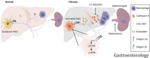
We identified cathepsin S (CTSS) up-regulation as a central node for extracellular matrix remodeling in the human fibrotic liver by proteomic screening. Increased serum CTSS levels efficiently predicted liver fibrosis, even at an early stage. Secreted CTSS cleaved collagen 18A1 at its C-terminus, releasing endostatin peptide, which directly bound to and activated hepatic stellate cells via integrin α5β1 signaling, whereas genetic ablation of Ctss remarkably suppressed liver fibrogenesis via endostatin reduction in vivo. Further studies identified macrophages as the main source of hepatic CTSS, and splenectomy effectively attenuated macrophage infiltration and CTSS expression in the fibrotic liver. Pharmacologic inhibition of CTSS ameliorated liver fibrosis progression in the mouse models.
Conclusions
CTSS functions as a novel profibrotic factor by remodeling extracellular matrix proteins and may represent a promising target for the diagnosis and treatment of liver fibrosis.
中文翻译:
巨噬细胞衍生的组织蛋白酶 S 重塑细胞外基质促进肝纤维化
背景与目标
肝纤维化是对慢性损伤的内在伤口愈合反应,也是全世界肝脏相关发病率和死亡率的主要原因。然而,由于其分子病因学特征尚不清楚,因此没有有效的诊断或治疗策略。我们的目的是阐明肝纤维形成的机制。
方法
我们对临床纤维化肝脏样本进行了定量蛋白质组分析,以识别失调的蛋白质。对 164 名肝纤维化患者的血清进行了进一步分析。使用两种纤维化小鼠模型和一些生化实验来阐明肝纤维化。
结果
我们通过蛋白质组学筛选,确定组织蛋白酶 S (CTSS) 上调是人纤维化肝脏细胞外基质重塑的中心节点。血清 CTSS 水平升高可以有效预测肝纤维化,即使是在早期阶段。分泌的 CTSS 在其 C 末端裂解胶原蛋白 18A1,释放内皮抑素肽,该肽通过整合素 α5β1 信号直接结合并激活肝星状细胞Ctss的基因消融则通过体内内皮抑素的减少显着抑制肝纤维化。进一步的研究发现巨噬细胞是肝脏CTSS的主要来源,脾切除可有效减弱纤维化肝脏中巨噬细胞的浸润和CTSS的表达。CTSS 的药理学抑制可改善小鼠模型中的肝纤维化进展。
结论
CTSS 通过重塑细胞外基质蛋白作为一种新型促纤维化因子发挥作用,可能是诊断和治疗肝纤维化的一个有希望的靶点。


 京公网安备 11010802027423号