当前位置:
X-MOL 学术
›
Anal. Chem.
›
论文详情
Our official English website, www.x-mol.net, welcomes your feedback! (Note: you will need to create a separate account there.)
Correction to “Effect of Exogenous Electric Stimulation on the Cardiac Tissue Function In Situ Monitored by Scanning Electrochemical Microscopy”
Analytical Chemistry ( IF 7.4 ) Pub Date : 2023-05-23 , DOI: 10.1021/acs.analchem.3c02009 Zhaoyang Ye , Yabei Li , Yuxiang Zhao , Junjie Zhang , Tong Zhu , Feng Xu , Fei Li
Analytical Chemistry ( IF 7.4 ) Pub Date : 2023-05-23 , DOI: 10.1021/acs.analchem.3c02009 Zhaoyang Ye , Yabei Li , Yuxiang Zhao , Junjie Zhang , Tong Zhu , Feng Xu , Fei Li
|
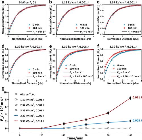
The authors would like to correct the unit of the vertical axis in Figure 5g of the original article (https://pubs.acs.org/doi/10.1021/acs.analchem.2c04758), in which the unit of Pm should be “m s–1” instead of “μm s–1”. The corrected Figure 5 is shown below. This correction does not affect the interpretation of the data or the conclusion of the article. The authors apologize for any inconvenience this might have caused. Figure 5. SECM experimental approaching curves and theoretical approaching curves to cardiac tissues after electric field stimulations with electric field strengths and electrical energies of (a) 0 kV cm–1, 0 J, (b) 1.19 kV cm–1, 0.001 J, (c) 2.37 kV cm–1, 0.001 J, (d) 3.39 kV cm–1, 0.001 J, (e) 3.39 kV cm–1, 0.005 J, and (f) 3.39 kV cm–1, 0.011 J. (g) Relationship between the average Pm values of cardiac tissues and the time after electric field stimulations (n = 3). SECM experiments were performed in a L15 solution containing 1 mM [Ru(NH3)6]Cl3 with approaching speeds of 0.5 μm s–1 and Eprobe of −0.35 V (vs Ag/AgCl RE). This article has not yet been cited by other publications. Figure 5. SECM experimental approaching curves and theoretical approaching curves to cardiac tissues after electric field stimulations with electric field strengths and electrical energies of (a) 0 kV cm–1, 0 J, (b) 1.19 kV cm–1, 0.001 J, (c) 2.37 kV cm–1, 0.001 J, (d) 3.39 kV cm–1, 0.001 J, (e) 3.39 kV cm–1, 0.005 J, and (f) 3.39 kV cm–1, 0.011 J. (g) Relationship between the average Pm values of cardiac tissues and the time after electric field stimulations (n = 3). SECM experiments were performed in a L15 solution containing 1 mM [Ru(NH3)6]Cl3 with approaching speeds of 0.5 μm s–1 and Eprobe of −0.35 V (vs Ag/AgCl RE).
中文翻译:

更正“外源性电刺激对扫描电化学显微镜原位监测心脏组织功能的影响”
作者想更正原文(https://pubs.acs.org/doi/10.1021/acs.analchem.2c04758)图5g中纵轴的单位,其中P m的单位应该是“ms –1 ”而不是“μm s –1 ”。修正后的图 5 如下所示。此更正不影响对数据的解释或文章的结论。对于由此可能造成的任何不便,作者深表歉意。图 5. 电场强度和电能为 (a) 0 kV cm –1 , 0 J , (b) 1.19 kV cm –1 , 0.001 J的电场刺激后心脏组织的 SECM 实验接近曲线和理论接近曲线(c) 2.37 千伏厘米–1, 0.001 J, (d) 3.39 kV cm –1 , 0.001 J, (e) 3.39 kV cm –1 , 0.005 J, (f) 3.39 kV cm –1 , 0.011 J。 (g) 平均P m之间的关系心脏组织的值和电场刺激后的时间 ( n = 3)。SECM 实验在含有 1 mM [Ru(NH 3 ) 6 ]Cl 3的 L15 溶液中进行,接近速度为 0.5 μm s –1,E探针-0.35 V(相对于 Ag/AgCl RE)。这篇文章尚未被其他出版物引用。图 5. 电场强度和电能为 (a) 0 kV cm –1 , 0 J , (b) 1.19 kV cm –1 , 0.001 J的电场刺激后心脏组织的 SECM 实验接近曲线和理论接近曲线(c) 2.37 kV cm –1 , 0.001 J, (d) 3.39 kV cm –1 , 0.001 J, (e) 3.39 kV cm –1 , 0.005 J, (f) 3.39 kV cm –1 , 0.011 J. ( g) 心肌组织平均P m值与电场刺激后时间的关系( n= 3). SECM 实验在含有 1 mM [Ru(NH 3 ) 6 ]Cl 3的 L15 溶液中进行,接近速度为 0.5 μm s –1,E探针为 −0.35 V(相对于 Ag/AgCl RE)。
更新日期:2023-05-23
中文翻译:
更正“外源性电刺激对扫描电化学显微镜原位监测心脏组织功能的影响”
作者想更正原文(https://pubs.acs.org/doi/10.1021/acs.analchem.2c04758)图5g中纵轴的单位,其中P m的单位应该是“ms –1 ”而不是“μm s –1 ”。修正后的图 5 如下所示。此更正不影响对数据的解释或文章的结论。对于由此可能造成的任何不便,作者深表歉意。图 5. 电场强度和电能为 (a) 0 kV cm –1 , 0 J , (b) 1.19 kV cm –1 , 0.001 J的电场刺激后心脏组织的 SECM 实验接近曲线和理论接近曲线(c) 2.37 千伏厘米–1, 0.001 J, (d) 3.39 kV cm –1 , 0.001 J, (e) 3.39 kV cm –1 , 0.005 J, (f) 3.39 kV cm –1 , 0.011 J。 (g) 平均P m之间的关系心脏组织的值和电场刺激后的时间 ( n = 3)。SECM 实验在含有 1 mM [Ru(NH 3 ) 6 ]Cl 3的 L15 溶液中进行,接近速度为 0.5 μm s –1,E探针-0.35 V(相对于 Ag/AgCl RE)。这篇文章尚未被其他出版物引用。图 5. 电场强度和电能为 (a) 0 kV cm –1 , 0 J , (b) 1.19 kV cm –1 , 0.001 J的电场刺激后心脏组织的 SECM 实验接近曲线和理论接近曲线(c) 2.37 kV cm –1 , 0.001 J, (d) 3.39 kV cm –1 , 0.001 J, (e) 3.39 kV cm –1 , 0.005 J, (f) 3.39 kV cm –1 , 0.011 J. ( g) 心肌组织平均P m值与电场刺激后时间的关系( n= 3). SECM 实验在含有 1 mM [Ru(NH 3 ) 6 ]Cl 3的 L15 溶液中进行,接近速度为 0.5 μm s –1,E探针为 −0.35 V(相对于 Ag/AgCl RE)。


 京公网安备 11010802027423号