Nature Materials ( IF 37.2 ) Pub Date : 2023-05-22 , DOI: 10.1038/s41563-023-01558-5 Kemal Arda Günay 1 , Tze-Ling Chang 1 , Nathaniel P Skillin 1 , Varsha V Rao 1 , Laura J Macdougall 1 , Alicia A Cutler 2 , Jason S Silver 1, 2 , Tobin E Brown 1 , Chi Zhang 3, 4, 5 , Chih-Chieh Jay Yu 3, 4, 5 , Bradley B Olwin 2 , Edward S Boyden 3, 4, 5 , Kristi S Anseth 1
|
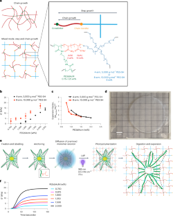
Hydrogels are extensively used as tunable, biomimetic three-dimensional cell culture matrices, but optically deep, high-resolution images are often difficult to obtain, limiting nanoscale quantification of cell–matrix interactions and outside-in signalling. Here we present photopolymerized hydrogels for expansion microscopy that enable optical clearance and tunable ×4.6–6.7 homogeneous expansion of not only monolayer cell cultures and tissue sections, but cells embedded within hydrogels. The photopolymerized hydrogels for expansion microscopy formulation relies on a rapid photoinitiated thiol/acrylate mixed-mode polymerization that is not inhibited by oxygen and decouples monomer diffusion from polymerization, which is particularly beneficial when expanding cells embedded within hydrogels. Using this technology, we visualize human mesenchymal stem cells and their interactions with nascently deposited proteins at <120 nm resolution when cultured in proteolytically degradable synthetic polyethylene glycol hydrogels. Results support the notion that focal adhesion maturation requires cellular fibronectin deposition; nuclear deformation precedes cellular spreading; and human mesenchymal stem cells display cell-surface metalloproteinases for matrix remodelling.
中文翻译:
光膨胀显微镜能够对嵌入 3D 水凝胶中的细胞进行超分辨率成像
水凝胶被广泛用作可调谐、仿生三维细胞培养基质,但光学深度、高分辨率图像通常难以获得,限制了细胞-基质相互作用和由外向内信号传导的纳米级量化。在这里,我们提出了用于膨胀显微镜的光聚合水凝胶,不仅可以实现单层细胞培养物和组织切片的光学间隙和可调×4.6-6.7均匀膨胀,还可以实现嵌入水凝胶中的细胞的光学间隙和可调×4.6-6.7均匀膨胀。用于膨胀显微镜制剂的光聚合水凝胶依赖于快速光引发硫醇/丙烯酸酯混合模式聚合,该聚合不受氧气抑制,并且使单体扩散与聚合解耦,这在膨胀嵌入水凝胶内的细胞时特别有益。利用这项技术,我们在可蛋白水解降解的合成聚乙二醇水凝胶中培养时,以 <120 nm 的分辨率可视化人类间充质干细胞及其与新生沉积蛋白质的相互作用。结果支持粘着斑成熟需要细胞纤连蛋白沉积的观点;核变形先于细胞扩散;人类间充质干细胞展示用于基质重塑的细胞表面金属蛋白酶。


 京公网安备 11010802027423号