Journal of Bioenergetics and Biomembranes ( IF 3 ) Pub Date : 2023-05-23 , DOI: 10.1007/s10863-023-09969-4 Miwa Takai 1 , Aya Okuda 1 , Yuka Amano 1 , Narumi Yashiro 1 , Koki Hara 1 , Mao Yamamoto 1 , Toshifumi Tsujiuchi 1
|
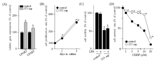
The tumor microenvironment (TME) consists of various cell types, including fibroblasts. The TME plays a central role in the promotion of tumor progression. In the present study, we investigated whether lysophosphatidic acid (LPA) receptor-mediated signaling regulates cellular functions by the TME of pancreatic cancer PANC-1 cells. To obtain fibroblast 3T3 cell supernatants, 3T3 cells were cultured in 5% charcoal stripped FCS-DMEM for 48 h. LPAR2 and LPAR3 expression levels were elevated in PANC-1 cells cultured in 3T3 cell supernatants. While PANC-1 cell motility was decreased by 3T3 cell supernatants, the cell survival to cisplatin (CDDP) of PANC-1 cells was markedly enhanced. Moreover, the cell survival to CDDP of PANC-1 cells cultured in 3T3 cell supernatants was increased by GRI-977,143 (LPA2 agonist) and (2 S)-OMPT (LPA3 agonist). Since hypoxia is caused by the restriction of adequate vascular networks to deliver oxygen into solid tumors, PANC-1 cells were cultured in 3T3 cell supernatants at 1% O2 conditions. The cell survival to CDDP of PANC-1 cells cultured in 3T3 cell supernatants at 1% O2 was significantly elevated, correlating with LPAR2 and LPAR3 expressions. These results suggest that LPA signaling via LPA2 and LPA3 is involved in the promotion of malignant properties by the TME in PANC-1 cells.
中文翻译:
LPA受体介导的信号传导对缺氧条件下成纤维细胞上清液中培养的胰腺癌细胞细胞功能调节的影响
肿瘤微环境(TME)由各种细胞类型组成,包括成纤维细胞。TME 在促进肿瘤进展中发挥着核心作用。在本研究中,我们研究了溶血磷脂酸 (LPA) 受体介导的信号传导是否通过胰腺癌 PANC-1 细胞的 TME 调节细胞功能。为了获得成纤维细胞3T3细胞上清液,将3T3细胞在5%木炭剥离的FCS-DMEM中培养48小时。在 3T3 细胞上清液中培养的 PANC-1 细胞中, LPAR2和LPAR3表达水平升高。虽然3T3细胞上清液降低了PANC-1细胞的运动性,但PANC-1细胞对顺铂(CDDP)的存活率却显着增强。此外,GRI-977,143(LPA 2激动剂)和(2 S)-OMPT(LPA 3激动剂)增加了在3T3细胞上清液中培养的PANC-1细胞对CDDP的细胞存活率。由于缺氧是由于将氧气输送到实体瘤的足够血管网络受到限制而引起的,因此在1% O 2条件下在3T3细胞上清液中培养PANC-1细胞。在 1% O 2的 3T3 细胞上清液中培养的 PANC-1 细胞的 CDDP 细胞存活率显着升高,与LPAR2和LPAR3表达相关。这些结果表明,通过 LPA 2和 LPA 3的 LPA 信号传导参与了 PANC-1 细胞中 TME 的恶性特性促进。


 京公网安备 11010802027423号