Nature ( IF 50.5 ) Pub Date : 2023-05-17 , DOI: 10.1038/s41586-023-05991-z Morris Degen 1 , José Carlos Santos 2 , Kristyna Pluhackova 3 , Gonzalo Cebrero 1 , Saray Ramos 2 , Gytis Jankevicius 1 , Ella Hartenian 2 , Undina Guillerm 4 , Stefania A Mari 5 , Bastian Kohl 1 , Daniel J Müller 5 , Paul Schanda 4 , Timm Maier 1 , Camilo Perez 1 , Christian Sieben 6, 7 , Petr Broz 2 , Sebastian Hiller 1
|
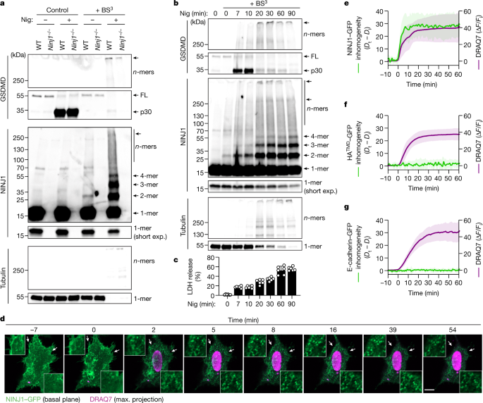
Eukaryotic cells can undergo different forms of programmed cell death, many of which culminate in plasma membrane rupture as the defining terminal event1,2,3,4,5,6,7. Plasma membrane rupture was long thought to be driven by osmotic pressure, but it has recently been shown to be in many cases an active process, mediated by the protein ninjurin-18 (NINJ1). Here we resolve the structure of NINJ1 and the mechanism by which it ruptures membranes. Super-resolution microscopy reveals that NINJ1 clusters into structurally diverse assemblies in the membranes of dying cells, in particular large, filamentous assemblies with branched morphology. A cryo-electron microscopy structure of NINJ1 filaments shows a tightly packed fence-like array of transmembrane α-helices. Filament directionality and stability is defined by two amphipathic α-helices that interlink adjacent filament subunits. The NINJ1 filament features a hydrophilic side and a hydrophobic side, and molecular dynamics simulations show that it can stably cap membrane edges. The function of the resulting supramolecular arrangement was validated by site-directed mutagenesis. Our data thus suggest that, during lytic cell death, the extracellular α-helices of NINJ1 insert into the plasma membrane to polymerize NINJ1 monomers into amphipathic filaments that rupture the plasma membrane. The membrane protein NINJ1 is therefore an interactive component of the eukaryotic cell membrane that functions as an in-built breaking point in response to activation of cell death.
中文翻译:
NINJ1介导的细胞死亡中质膜破裂的结构基础
真核细胞可以经历不同形式的程序性细胞死亡,其中许多最终导致质膜破裂作为定义的终止事件1,2,3,4,5,6,7 。长期以来,质膜破裂被认为是由渗透压驱动的,但最近表明,在许多情况下,质膜破裂是一个主动过程,由蛋白质 ninjurin-1 8 (NINJ1) 介导。在这里,我们解析了 NINJ1 的结构及其破膜机制。超分辨率显微镜显示,NINJ1 在垂死细胞膜中聚集成结构多样的组件,特别是具有分支形态的大型丝状组件。 NINJ1 细丝的冷冻电子显微镜结构显示出紧密排列的栅栏状跨膜 α 螺旋阵列。细丝的方向性和稳定性由两个连接相邻细丝亚基的两亲性 α 螺旋定义。 NINJ1丝具有亲水侧和疏水侧,分子动力学模拟表明它可以稳定地覆盖膜边缘。通过定点诱变验证了所得超分子排列的功能。因此,我们的数据表明,在裂解细胞死亡过程中,NINJ1 的细胞外 α-螺旋插入质膜,将 NINJ1 单体聚合成两亲丝,从而破裂质膜。因此,膜蛋白 NINJ1 是真核细胞膜的相互作用成分,作为响应细胞死亡激活的内置断裂点。


 京公网安备 11010802027423号