当前位置:
X-MOL 学术
›
J. Biophotonics
›
论文详情
Our official English website, www.x-mol.net, welcomes your
feedback! (Note: you will need to create a separate account there.)
Optical time-stretch imaging flow cytometry in the compressed domain
Journal of Biophotonics ( IF 2.0 ) Pub Date : 2023-05-11 , DOI: 10.1002/jbio.202300096 Siyuan Lin 1 , Rubing Li 1 , Yueyun Weng 1, 2 , Liye Mei 1 , Chao Wei 1 , Congkuan Song 3 , Shubin Wei 1 , Yifan Yao 1 , Xiaolan Ruan 4 , Fuling Zhou 5 , Qing Geng 3 , Du Wang 1 , Cheng Lei 1, 6
Journal of Biophotonics ( IF 2.0 ) Pub Date : 2023-05-11 , DOI: 10.1002/jbio.202300096 Siyuan Lin 1 , Rubing Li 1 , Yueyun Weng 1, 2 , Liye Mei 1 , Chao Wei 1 , Congkuan Song 3 , Shubin Wei 1 , Yifan Yao 1 , Xiaolan Ruan 4 , Fuling Zhou 5 , Qing Geng 3 , Du Wang 1 , Cheng Lei 1, 6
Affiliation
|
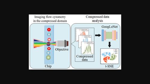
Imaging flow cytometry based on optical time-stretch (OTS) imaging combined with a microfluidic chip attracts much attention in the large-scale single-cell analysis due to its high throughput, high precision, and label-free operation. Compressive sensing has been integrated into OTS imaging to relieve the pressure on the sampling and transmission of massive data. However, image decompression brings an extra overhead of computing power to the system, but does not generate additional information. In this work, we propose and demonstrate OTS imaging flow cytometry in the compressed domain. Specifically, we constructed a machine-learning network to analyze the cells without decompressing the images. The results show that our system enables high-quality imaging and high-accurate cell classification with an accuracy of over 99% at a compression ratio of 10%. This work provides a viable solution to the big data problem in OTS imaging flow cytometry, boosting its application in practice.
中文翻译:

压缩域中的光学时间拉伸成像流式细胞术
基于光学时间拉伸(OTS)成像与微流控芯片相结合的成像流式细胞术因其高通量、高精度和免标记操作在大规模单细胞分析中备受关注。压缩感知已融入OTS成像,缓解海量数据采样和传输的压力。然而图像解压缩给系统带来了额外的计算能力开销,但不会产生额外的信息。在这项工作中,我们提出并演示了压缩域中的 OTS 成像流式细胞术。具体来说,我们构建了一个机器学习网络来分析细胞,而无需解压缩图像。结果表明,我们的系统能够实现高质量成像和高精度细胞分类,在10%的压缩比下,准确率超过99%。这项工作为OTS成像流式细胞术中的大数据问题提供了可行的解决方案,促进了其在实践中的应用。
更新日期:2023-05-11
中文翻译:
压缩域中的光学时间拉伸成像流式细胞术
基于光学时间拉伸(OTS)成像与微流控芯片相结合的成像流式细胞术因其高通量、高精度和免标记操作在大规模单细胞分析中备受关注。压缩感知已融入OTS成像,缓解海量数据采样和传输的压力。然而图像解压缩给系统带来了额外的计算能力开销,但不会产生额外的信息。在这项工作中,我们提出并演示了压缩域中的 OTS 成像流式细胞术。具体来说,我们构建了一个机器学习网络来分析细胞,而无需解压缩图像。结果表明,我们的系统能够实现高质量成像和高精度细胞分类,在10%的压缩比下,准确率超过99%。这项工作为OTS成像流式细胞术中的大数据问题提供了可行的解决方案,促进了其在实践中的应用。


 京公网安备 11010802027423号