Nature Nanotechnology ( IF 38.1 ) Pub Date : 2023-04-10 , DOI: 10.1038/s41565-023-01328-z Dominik Kylies 1, 2, 3 , Marina Zimmermann 1, 2, 4 , Fabian Haas 1, 2 , Maria Schwerk 1, 2 , Malte Kuehl 1, 2, 4 , Michael Brehler 4 , Jan Czogalla 1, 2 , Lola C Hernandez 5 , Leonie Konczalla 6, 7 , Yusuke Okabayashi 1, 2, 8 , Julia Menzel 9 , Ilka Edenhofer 1, 2 , Sam Mezher 1, 2 , Hande Aypek 1, 2 , Bernhard Dumoulin 1, 2 , Hui Wu 1, 2 , Smilla Hofmann 1, 2 , Oliver Kretz 1, 2 , Nicola Wanner 1, 2 , Nicola M Tomas 1, 2 , Susanne Krasemann 10 , Markus Glatzel 10 , Christoph Kuppe 11 , Rafael Kramann 11 , Bella Banjanin 12, 13 , Rebekka K Schneider 12, 13, 14 , Christopher Urbschat 15 , Petra Arck 15 , Nicola Gagliani 6, 16 , Marc van Zandvoort 17, 18 , Thorsten Wiech 19 , Florian Grahammer 1, 2 , Pablo J Sáez 5 , Milagros N Wong 1, 2, 20, 21 , Stefan Bonn 4 , Tobias B Huber 1, 2 , Victor G Puelles 1, 2, 20, 21
|
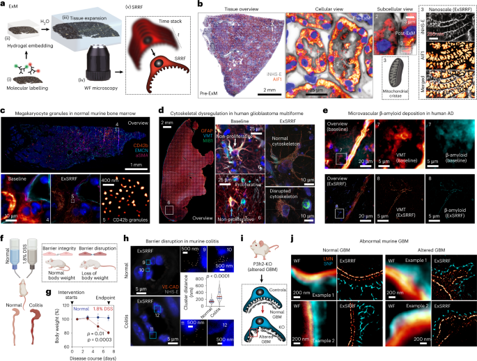
Expansion microscopy physically enlarges biological specimens to achieve nanoscale resolution using diffraction-limited microscopy systems1. However, optimal performance is usually reached using laser-based systems (for example, confocal microscopy), restricting its broad applicability in clinical pathology, as most centres have access only to light-emitting diode (LED)-based widefield systems. As a possible alternative, a computational method for image resolution enhancement, namely, super-resolution radial fluctuations (SRRF)2,3, has recently been developed. However, this method has not been explored in pathology specimens to date, because on its own, it does not achieve sufficient resolution for routine clinical use. Here, we report expansion-enhanced super-resolution radial fluctuations (ExSRRF), a simple, robust, scalable and accessible workflow that provides a resolution of up to 25 nm using LED-based widefield microscopy. ExSRRF enables molecular profiling of subcellular structures from archival formalin-fixed paraffin-embedded tissues in complex clinical and experimental specimens, including ischaemic, degenerative, neoplastic, genetic and immune-mediated disorders. Furthermore, as examples of its potential application to experimental and clinical pathology, we show that ExSRRF can be used to identify and quantify classical features of endoplasmic reticulum stress in the murine ischaemic kidney and diagnostic ultrastructural features in human kidney biopsies.
中文翻译:
扩展增强的超分辨率径向波动使病理标本的纳米级分子分析成为可能
膨胀显微镜使用衍射极限显微镜系统1物理放大生物标本以实现纳米级分辨率。然而,通常使用基于激光的系统(例如,共聚焦显微镜)可以达到最佳性能,这限制了它在临床病理学中的广泛适用性,因为大多数中心只能使用基于发光二极管 (LED) 的宽场系统。作为一种可能的替代方法,一种用于图像分辨率增强的计算方法,即超分辨率径向波动 (SRRF) 2,3,最近被开发出来。然而,迄今为止,这种方法还没有在病理标本中进行过探索,因为它本身并没有达到常规临床使用的足够分辨率。在这里,我们报告了扩展增强的超分辨率径向波动 (ExSRRF),这是一种简单、稳健、可扩展且易于访问的工作流程,使用基于 LED 的宽场显微镜可提供高达 25 nm 的分辨率。ExSRRF 能够对复杂临床和实验标本中福尔马林固定石蜡包埋组织的亚细胞结构进行分子分析,包括缺血、退行性、肿瘤、遗传和免疫介导的疾病。此外,作为其在实验和临床病理学中潜在应用的例子,


 京公网安备 11010802027423号