Scientific Reports ( IF 3.8 ) Pub Date : 2023-03-17 , DOI: 10.1038/s41598-023-31712-7 Rong Du 1 , Xi Cheng 1 , Jingjing Ji 1 , Yang Lu 2 , Yuanyuan Xie 1 , Weina Wang 1 , Yanhua Xu 1 , Yuquan Zhang 3
|
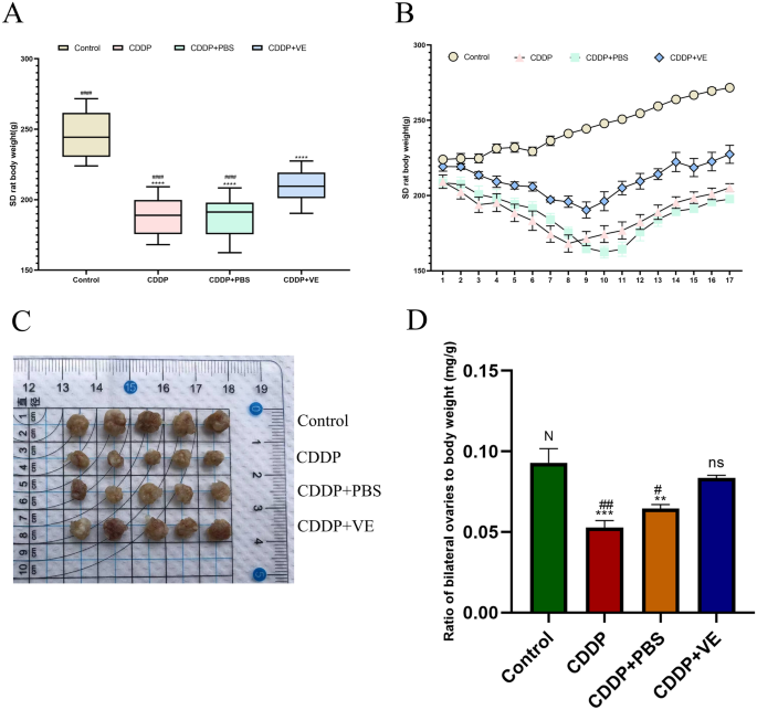
Ferroptosis is widely present in fibrosis-related diseases. The basic pathology of premature ovarian insufficiency (POI) involves ovarian tissue fibrosis, and there are currently fewer relevant studies addressing the association between ferroptosis and POI. This study aimed to demonstrate that ferroptosis induced by cisplatin (CDDP) caused ovarian tissue fibrosis, leading to POI. Vitamin E (VE), a ferroptosis inhibitor, could repair damaged ovarian function. CDDP was used to establish a rat model of POI, and VE was administered to reverse the reproductive toxicity of CDDP. Ovarian function was assessed by histological section staining, follicle counts, sex hormone levels, as well as fertility assays. The extent of ferroptosis was assessed by transmission electron microscopy (TEM), malondialdehyde (MDA), Perls staining. CCK-8, Ethynyl-2-Deoxyuridine (EdU), and scratch assays were used to determine the effect of CDDP and VE on ovarian granulosa cell (GC) viability. Western blot, quantitative reverse-transcription polymerase chain reaction (qRT-PCR) and immunohistochemistry were performed to evaluate ferroptosis-related molecular changes. Our results showed that CDDP caused follicle development disorders and ovarian tissue fibrosis, the levels of sex hormones suggested impaired ovarian function, and VE could reverse the reproductive toxicity of CDDP. The results of TEM, MDA and Perls staining suggested that the typical mitochondrial signature of ferroptosis was altered in ovarian GCs from the CDDP group, with significantly higher levels of lipid peroxidation and significant iron deposition in ovarian tissue, whereas VE mitigated the extent of ferroptosis. Molecular experiments then confirmed that the ferroptosis-related molecules acetyl CoA synthetase long chain family member 4 (ACSl4), 15-lipoxygenase-1 (ALOX15), solute carrier family 7 member 11 (SLC7A11), and glutathione peroxidase 4 (GPX4) were differentially expressed in each group. In summary, our study preliminarily demonstrated that CDDP may promote GCs to undergo ferroptosis, cause follicle development disorders, ovarian tissue fibrosis, and induce POI by regulating the expression of ACSl4, ALOX15, SLC7A11, and GPX4, while VE improved impaired ovarian function.
中文翻译:
顺铂致卵巢早衰模型大鼠铁死亡机制
铁死亡广泛存在于纤维化相关疾病中。早产卵巢功能不全(POI)的基本病理学涉及卵巢组织纤维化,目前针对铁死亡与POI之间关系的相关研究较少。本研究旨在证明顺铂 (CDDP) 诱导的铁死亡导致卵巢组织纤维化,从而导致 POI。维生素 E (VE) 是一种铁死亡抑制剂,可以修复受损的卵巢功能。采用CDDP建立POI大鼠模型,给予VE逆转CDDP的生殖毒性。卵巢功能通过组织切片染色、卵泡计数、性激素水平以及生育力测定来评估。通过透射电子显微镜 (TEM)、丙二醛 (MDA)、Perls 染色评估铁死亡的程度。CCK-8, 乙炔基-2-脱氧尿苷 (EdU) 和划痕试验用于确定 CDDP 和 VE 对卵巢颗粒细胞 (GC) 活力的影响。进行蛋白质印迹、定量逆转录聚合酶链反应 (qRT-PCR) 和免疫组织化学以评估铁死亡相关的分子变化。我们的研究结果表明,CDDP引起卵泡发育障碍和卵巢组织纤维化,性激素水平提示卵巢功能受损,VE可以逆转CDDP的生殖毒性。TEM、MDA 和 Perls 染色的结果表明,CDDP 组卵巢 GC 中铁死亡的典型线粒体特征发生了改变,卵巢组织中脂质过氧化水平显着升高,铁沉积显着,而 VE 减轻了铁死亡的程度。分子实验随后证实,与铁死亡相关的分子乙酰辅酶 A 合成酶长链家族成员 4 (ACSl4)、15-脂氧合酶-1 (ALOX15)、溶质载体家族 7 成员 11 (SLC7A11) 和谷胱甘肽过氧化物酶 4 (GPX4) 存在差异表达在每组中。综上所述,本研究初步证明CDDP可能通过调节ACSl4、ALOX15、SLC7A11、GPX4的表达促进GC发生铁死亡、卵泡发育障碍、卵巢组织纤维化、POI,而VE改善卵巢功能受损。


 京公网安备 11010802027423号