Nature Communications ( IF 14.7 ) Pub Date : 2022-11-30 , DOI: 10.1038/s41467-022-35026-6 Xia Yao 1 , Yan Wang 2 , Zhifei Wang 2 , Xiao Fan 1 , Di Wu 3, 4 , Jian Huang 1 , Alexander Mueller 1 , Sarah Gao 1 , Miaohui Hu 1 , Carol V Robinson 3, 4 , Yong Yu 2 , Shuai Gao 1, 5 , Nieng Yan 1
|
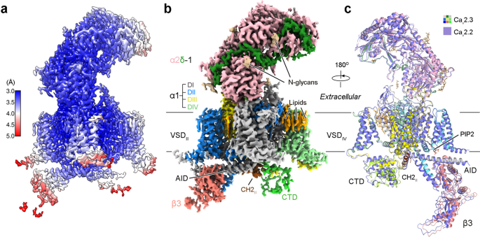
The R-type voltage-gated Ca2+ (Cav) channels Cav2.3, widely expressed in neuronal and neuroendocrine cells, represent potential drug targets for pain, seizures, epilepsy, and Parkinson’s disease. Despite their physiological importance, there have lacked selective small-molecule inhibitors targeting these channels. High-resolution structures may aid rational drug design. Here, we report the cryo-EM structure of human Cav2.3 in complex with α2δ−1 and β3 subunits at an overall resolution of 3.1 Å. The structure is nearly identical to that of Cav2.2, with VSDII in the down state and the other three VSDs up. A phosphatidylinositol 4,5-bisphosphate (PIP2) molecule binds to the interface of VSDII and the tightly closed pore domain. We also determined the cryo-EM structure of a Cav2.3 mutant in which a Cav2-unique cytosolic helix in repeat II (designated the CH2II helix) is deleted. This mutant, named ΔCH2, still reserves a down VSDII, but PIP2 is invisible and the juxtamembrane region on the cytosolic side is barely discernible. Our structural and electrophysiological characterizations of the wild type and ΔCH2 Cav2.3 show that the CH2II helix stabilizes the inactivated conformation of the channel by tightening the cytosolic juxtamembrane segments, while CH2II helix is not necessary for locking the down state of VSDII.
中文翻译:
R 型人 Cav2.3 通道的结构揭示了细胞内片段的构象串扰
R 型电压门控 Ca 2+ (Ca v ) 通道 Ca v 2.3 在神经元和神经内分泌细胞中广泛表达,是治疗疼痛、癫痫、癫痫和帕金森病的潜在药物靶点。尽管它们具有生理重要性,但缺乏针对这些通道的选择性小分子抑制剂。高分辨率结构可能有助于合理的药物设计。在这里,我们报告了人类 Ca v 2.3 与 α2δ−1 和 β3 亚基复合物的低温电子显微镜结构,总分辨率为 3.1 Å。该结构与 Ca v 2.2 的结构几乎相同,具有 VSD II处于关闭状态,其他三个 VSD 处于关闭状态。磷脂酰肌醇 4,5-二磷酸 (PIP2) 分子结合到 VSD II的界面和紧密闭合的孔域。我们还确定了 Ca v 2.3 突变体的冷冻电镜结构,其中重复 II 中的 Ca v 2 独特胞质螺旋(指定为 CH2 II螺旋)被删除。这个突变体,命名为 ΔCH2,仍然保留下 VSD II,但 PIP2 是不可见的,胞质侧的近膜区域几乎看不到。我们对野生型和 ΔCH2 Ca v 2.3 的结构和电生理特征表明,CH2 II螺旋通过收紧胞质近膜区段来稳定通道的失活构象,而 CH2 II螺旋对于锁定 VSD II的向下状态不是必需的。


 京公网安备 11010802027423号