Our official English website, www.x-mol.net, welcomes your
feedback! (Note: you will need to create a separate account there.)
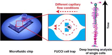
Effect of capillary fluid flow on single cancer cell cycle dynamics, motility, volume and morphology
Lab on a Chip ( IF 6.1 ) Pub Date : 2022-11-30 , DOI: 10.1039/d2lc00322h Hubert M Taïeb 1 , Guillaume Herment 1 , Tom Robinson 2 , Amaia Cipitria 1, 3, 4
Lab on a Chip ( IF 6.1 ) Pub Date : 2022-11-30 , DOI: 10.1039/d2lc00322h Hubert M Taïeb 1 , Guillaume Herment 1 , Tom Robinson 2 , Amaia Cipitria 1, 3, 4
Affiliation
|
From primary tu5mours and disseminating to secondary organs, cancer cells experience a wide variety of fluid flow profiles when passing through blood vessels or the lymphatic system before extravasation. Sinusoidal capillaries are a common site for extravasation. Therefore, we aim to investigate how metastatic cancer cells react to a biophysical cue such as capillary fluid flow by quantifying its effect on metastatic cell cycle progression, motility, cell and nuclear volume, and morphology. We use MDA-MB-231 breast cancer cells genetically modified with fluorescent ubiquitination-based cell cycle indicator 2 (FUCCI2) as a model system. Single cells are trapped using a microfluidic device and exposed to different laminar flows. Quantitative time-lapse imaging in both 2D epifluorescence and 3D confocal microscopy is performed using in-house software FUCCItrack. In addition, 3D time-lapse with cell and nuclear segmentation is performed with a deep learning approach to streamline the image processing of big datasets. We show that at a single cell level, faster fluid flow leads to a shorter S/G2/M phase and an overall shorter cell cycle, as well as increase in cell motility irrespective of the flow direction. 3D time-lapse confocal imaging of MDA-FUCCI2 single cells reveals the evolution of cell and nuclear volume and morphology as a function of a specific cell cycle phase. Both cell and nuclear volume increase linearly over time. Cell morphology elongates more strongly during the S/G2/M phase, whereas the nuclear shape remains constant. Under the highest flow conditions, only during the S/G2/M phase can we observe a more elongated nucleus, while the cell sphericity remains similar to the control. Collectively, this data, together with the deep learning approach, provides new insights into the potential impact of fluid flow at a single cell level.
中文翻译:

毛细管流体流动对单个癌细胞周期动力学、运动性、体积和形态的影响
从原发性肿瘤和扩散到次级器官,癌细胞在外渗前通过血管或淋巴系统时会经历各种各样的流体流动曲线。正弦曲线毛细血管是外渗的常见部位。因此,我们的目标是通过量化其对转移性细胞周期进程、运动性、细胞和核体积以及形态的影响来研究转移性癌细胞如何对毛细管流体流动等生物物理线索作出反应。我们使用基于荧光泛素化的细胞周期指示剂 2 (FUCCI2) 进行基因修饰的 MDA-MB-231 乳腺癌细胞作为模型系统。使用微流体装置捕获单个细胞并暴露于不同的层流。2D 落射荧光和 3D 共聚焦显微镜中的定量延时成像是使用内部软件 FUCCItrack 进行的。此外,使用深度学习方法执行细胞和核分割的 3D 延时,以简化大数据集的图像处理。我们表明,在单个细胞水平上,更快的流体流动会导致更短的 S/G2/M 期和更短的整体细胞周期,以及无论流动方向如何都会增加细胞运动性。MDA-FUCCI2 单细胞的 3D 延时共聚焦成像揭示了细胞和核体积和形态随特定细胞周期阶段的变化。细胞和核体积都随时间线性增加。在 S/G2/M 阶段,细胞形态更加强烈地伸长,而核形状保持不变。在最高流量条件下,只有在 S/G2/M 阶段,我们才能观察到更细长的细胞核,而细胞球形度与对照相似。总的来说,这些数据与深度学习方法一起,为了解流体流动在单个细胞水平上的潜在影响提供了新的见解。
更新日期:2022-11-30
中文翻译:
毛细管流体流动对单个癌细胞周期动力学、运动性、体积和形态的影响
从原发性肿瘤和扩散到次级器官,癌细胞在外渗前通过血管或淋巴系统时会经历各种各样的流体流动曲线。正弦曲线毛细血管是外渗的常见部位。因此,我们的目标是通过量化其对转移性细胞周期进程、运动性、细胞和核体积以及形态的影响来研究转移性癌细胞如何对毛细管流体流动等生物物理线索作出反应。我们使用基于荧光泛素化的细胞周期指示剂 2 (FUCCI2) 进行基因修饰的 MDA-MB-231 乳腺癌细胞作为模型系统。使用微流体装置捕获单个细胞并暴露于不同的层流。2D 落射荧光和 3D 共聚焦显微镜中的定量延时成像是使用内部软件 FUCCItrack 进行的。此外,使用深度学习方法执行细胞和核分割的 3D 延时,以简化大数据集的图像处理。我们表明,在单个细胞水平上,更快的流体流动会导致更短的 S/G2/M 期和更短的整体细胞周期,以及无论流动方向如何都会增加细胞运动性。MDA-FUCCI2 单细胞的 3D 延时共聚焦成像揭示了细胞和核体积和形态随特定细胞周期阶段的变化。细胞和核体积都随时间线性增加。在 S/G2/M 阶段,细胞形态更加强烈地伸长,而核形状保持不变。在最高流量条件下,只有在 S/G2/M 阶段,我们才能观察到更细长的细胞核,而细胞球形度与对照相似。总的来说,这些数据与深度学习方法一起,为了解流体流动在单个细胞水平上的潜在影响提供了新的见解。


 京公网安备 11010802027423号