Nature ( IF 64.8 ) Pub Date : 2022-11-23 , DOI: 10.1038/s41586-022-05449-8 Nisha Pillay 1, 2 , Laura Mariotti 1, 2 , Mariola Zaleska 1, 2 , Oviya Inian 1, 2 , Matthew Jessop 1, 2 , Sam Hibbs 1, 2 , Ambroise Desfosses 3 , Paul C R Hopkins 1, 2 , Catherine M Templeton 1, 2 , Fabienne Beuron 1 , Edward P Morris 1 , Sebastian Guettler 1, 2
|
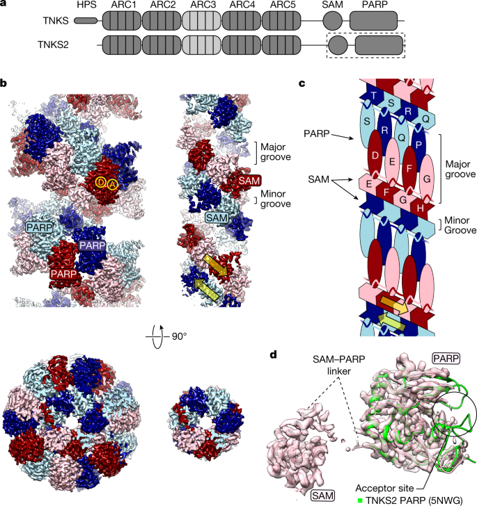
The poly-ADP-ribosyltransferase tankyrase (TNKS, TNKS2) controls a wide range of disease-relevant cellular processes, including WNT–β-catenin signalling, telomere length maintenance, Hippo signalling, DNA damage repair and glucose homeostasis1,2. This has incentivized the development of tankyrase inhibitors. Notwithstanding, our knowledge of the mechanisms that control tankyrase activity has remained limited. Both catalytic and non-catalytic functions of tankyrase depend on its filamentous polymerization3,4,5. Here we report the cryo-electron microscopy reconstruction of a filament formed by a minimal active unit of tankyrase, comprising the polymerizing sterile alpha motif (SAM) domain and its adjacent catalytic domain. The SAM domain forms a novel antiparallel double helix, positioning the protruding catalytic domains for recurring head-to-head and tail-to-tail interactions. The head interactions are highly conserved among tankyrases and induce an allosteric switch in the active site within the catalytic domain to promote catalysis. Although the tail interactions have a limited effect on catalysis, they are essential to tankyrase function in WNT–β-catenin signalling. This work reveals a novel SAM domain polymerization mode, illustrates how supramolecular assembly controls catalytic and non-catalytic functions, provides important structural insights into the regulation of a non-DNA-dependent poly-ADP-ribosyltransferase and will guide future efforts to modulate tankyrase and decipher its contribution to disease mechanisms.
中文翻译:
聚合激活端锚聚合酶的结构基础
聚-ADP-核糖基转移酶端锚聚合酶 (TNKS, TNKS2) 控制广泛的疾病相关细胞过程,包括 WNT-β-连环蛋白信号传导、端粒长度维持、Hippo 信号传导、DNA 损伤修复和葡萄糖稳态1,2。这刺激了端锚聚合酶抑制剂的开发。尽管如此,我们对控制端锚聚合酶活性的机制的了解仍然有限。tankyrase 的催化和非催化功能均取决于其丝状聚合3,4,5. 在这里,我们报告了由端锚聚合酶的最小活性单元形成的细丝的低温电子显微镜重建,包括聚合无菌 α 基序 (SAM) 结构域及其相邻的催化结构域。SAM 结构域形成一个新的反平行双螺旋,定位突出的催化结构域以进行重复的头对头和尾对尾相互作用。头部相互作用在端锚聚合酶中高度保守,并在催化域内的活性位点诱导变构开关以促进催化作用。尽管尾部相互作用对催化的影响有限,但它们对于 WNT-β-连环蛋白信号传导中的端锚聚合酶功能至关重要。这项工作揭示了一种新的 SAM 域聚合模式,说明了超分子组装如何控制催化和非催化功能,


 京公网安备 11010802027423号