Nature Nanotechnology ( IF 38.3 ) Pub Date : 2022-10-31 , DOI: 10.1038/s41565-022-01224-y Zhen Chen 1, 2 , Emrah Turgut 3 , Yi Jiang 4 , Kayla X Nguyen 5 , Matthew J Stolt 6 , Song Jin 6 , Daniel C Ralph 7, 8 , Gregory D Fuchs 1 , David A Muller 1, 8
|
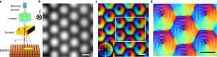
Nanoscale spin textures, especially magnetic skyrmions, have attracted intense interest as candidate high-density and power-efficient information carriers for spintronic devices1,2. Facilitating a deeper understanding of sub-hundred-nanometre to atomic-scale spin textures requires more advanced magnetic imaging techniques3,4,5. Here we demonstrate a Lorentz electron ptychography method that can enable high-resolution, high-sensitivity magnetic field imaging for widely available electron microscopes. The resolution of Lorentz electron ptychography is not limited by the usual diffraction limit of lens optics, but instead is determined by the maximum scattering angle at which a statistically meaningful dose can still be recorded—this can be an improvement of up to 2–6 times depending on the allowable dose. Using FeGe as the model system, we realize a more accurate magnetic field measurement of skyrmions with an improved spatial resolution and sensitivity by also correcting the probe-damping effect from the imaging optics via Lorentz electron ptychography. This allows us to directly resolve subtle internal structures of magnetic skyrmions near the skyrmion cores, boundaries and dislocations in an FeGe single crystal. Our study establishes a quantitative, high-resolution magnetic microscopy technique that can reveal nanoscale spin textures, especially magnetization discontinuities and topological defects in nanomagnets6. The technique’s high-dose efficiency should also make it well suited for the exploration of magnetic textures in electron radiation-sensitive materials such as organic or molecular magnets7.
中文翻译:
用于成像超出衍射极限的磁纹理的洛伦兹电子叠层描记法
纳米级自旋纹理,尤其是磁性斯格明子,作为自旋电子设备1,2的候选高密度和高能效信息载体,引起了人们的浓厚兴趣。促进对亚百纳米到原子级自旋纹理的更深入理解需要更先进的磁成像技术3,4,5. 在这里,我们展示了一种洛伦兹电子叠印法,该方法可以为广泛使用的电子显微镜实现高分辨率、高灵敏度的磁场成像。洛伦兹电子层积法的分辨率不受透镜光学器件通常的衍射极限的限制,而是由仍然可以记录具有统计意义的剂量的最大散射角决定——这可以提高 2-6 倍取决于允许的剂量。使用 FeGe 作为模型系统,我们通过洛伦兹电子叠层图校正成像光学的探针阻尼效应,实现了更准确的 skyrmions 磁场测量,提高了空间分辨率和灵敏度。这使我们能够直接解析靠近 skyrmion 核心的磁性 skyrmion 的微妙内部结构,FeGe 单晶中的晶界和位错。我们的研究建立了一种定量的高分辨率磁显微技术,可以揭示纳米级自旋纹理,尤其是纳米磁体中的磁化不连续性和拓扑缺陷6 . 该技术的高剂量效率还应该使其非常适合探索电子辐射敏感材料(例如有机或分子磁体7 )中的磁性结构。


 京公网安备 11010802027423号